Copyright
©2009 The WJG Press and Baishideng.
World J Gastroenterol. Nov 14, 2009; 15(42): 5326-5333
Published online Nov 14, 2009. doi: 10.3748/wjg.15.5326
Published online Nov 14, 2009. doi: 10.3748/wjg.15.5326
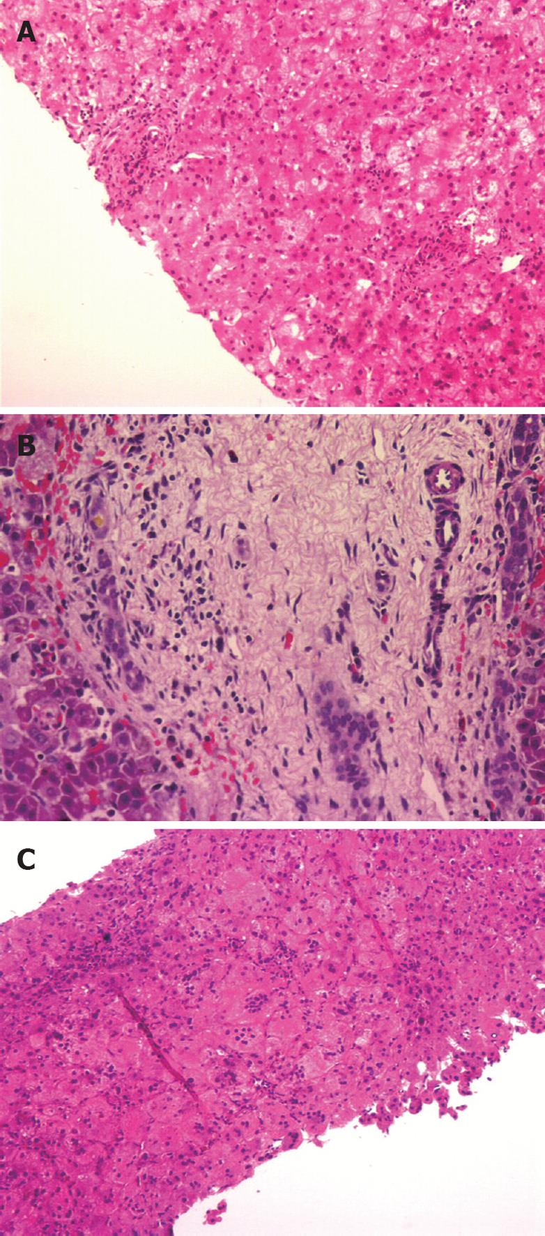
Figure 1 Photomicrograph of liver biopsy (hematoxylin and eosin, original magnification × 200).
A: A patient with biliary atresia obtained at 34 d of age (Patient NKC). A portal tract is shown here with no obvious bile ductular proliferation. There was hepatocytic degeneration and swelling. The original blinded histological diagnosis by the author was neonatal hepatitis; B: NKC obtained at 45 d of age. A portal tract is shown here with marked bile ductular proliferation, and bile plugs in bile ductules, typical of biliary atresia; C: A patient with biliary atresia (LQY) obtained at 78 d of life. There are multiple multinucleated giant cells, hepatocellular swelling and moderate lymphocytic infiltration. The original blinded histological diagnosis by the author was neonatal hepatitis.
- Citation: Lee WS, Looi LM. Usefulness of a scoring system in the interpretation of histology in neonatal cholestasis. World J Gastroenterol 2009; 15(42): 5326-5333
- URL: https://www.wjgnet.com/1007-9327/full/v15/i42/5326.htm
- DOI: https://dx.doi.org/10.3748/wjg.15.5326


