Copyright
©2009 The WJG Press and Baishideng.
World J Gastroenterol. Nov 7, 2009; 15(41): 5221-5223
Published online Nov 7, 2009. doi: 10.3748/wjg.15.5221
Published online Nov 7, 2009. doi: 10.3748/wjg.15.5221
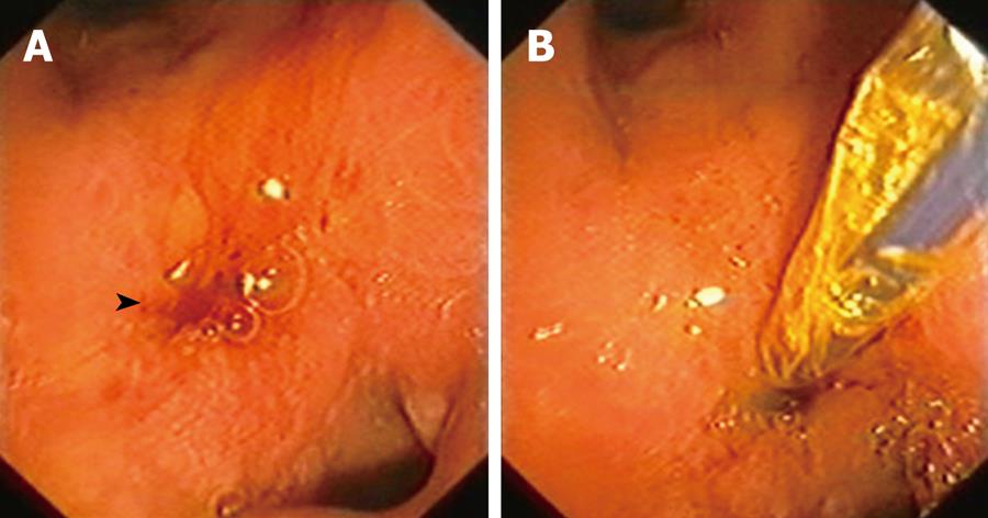
Figure 1 Endoscopic retrograde cholangiopancreatography.
A: View of the papilla located in the pylorus (arrow head); B: Piping of the papilla.
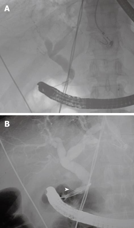
Figure 2 Cholangiography.
A: Common bile duct ending in a hook shape, common in proximal drainage of the papilla; B: Removal of choledocholithiasis with Dormia basket (arrow head).
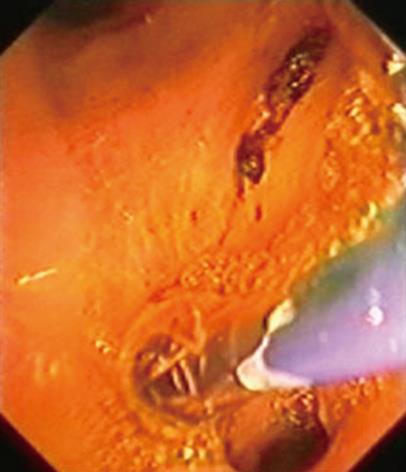
Figure 3 Endoscopic retrograde cholangiopancreatography.
Balloon dilatation of the ectopic papilla.
- Citation: Guerra I, Rábago LR, Bermejo F, Quintanilla E, García-Garzón S. Ectopic papilla of Vater in the pylorus. World J Gastroenterol 2009; 15(41): 5221-5223
- URL: https://www.wjgnet.com/1007-9327/full/v15/i41/5221.htm
- DOI: https://dx.doi.org/10.3748/wjg.15.5221


