Copyright
©2009 The WJG Press and Baishideng.
World J Gastroenterol. Oct 28, 2009; 15(40): 5028-5034
Published online Oct 28, 2009. doi: 10.3748/wjg.15.5028
Published online Oct 28, 2009. doi: 10.3748/wjg.15.5028
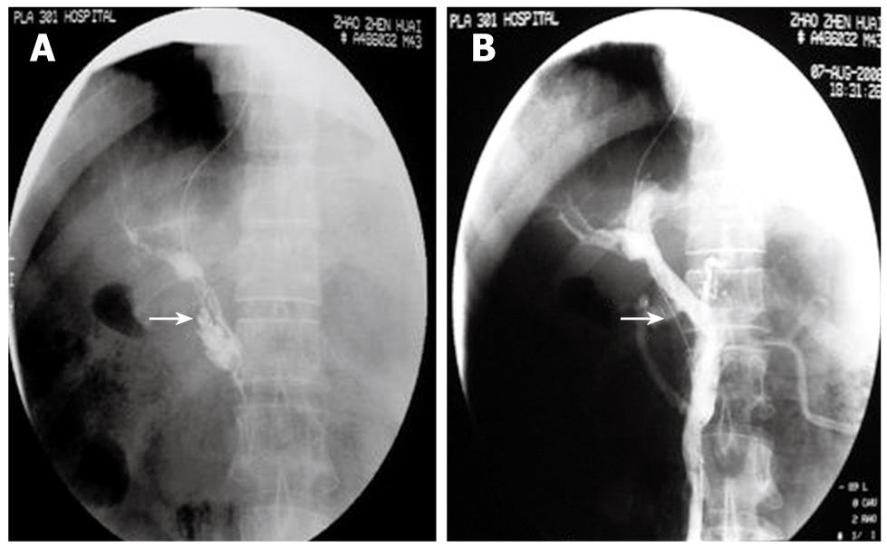
Figure 1 The figure shows a male, 43-year-old patient, 2 mo after splenectomy, with abdominal fullness for 6 d.
He had thrombolysis through a transjugular intrahepatic portosystemic shunt (TIPS). A: Direct PV-SMV angiography showed extensive thrombosis in the PV and SMV. Contrast agent remained. No lateral branch angiogenesis (arrow); B: Mash and suck thrombi with intermittent injection of urokinase and heparin sodium. Repeated angiography 30 min after thrombolysis showed blood reperfusion in the PV-SMV and normal PV-SMV branches (arrow). Symptoms disappeared.
Figure 2 The figure shows a female, 22 year old patient who had abdominal pain for 17 d.
A: A CT scan showed a high density of superior mesenteric vein thrombosis (characteristic of subacute thrombosis) (arrow); B: A Cobra catheter was inserted via the femoral artery into the superior mesenteric artery. Indirect angiography showed extensive thrombosis in the PV-SMV. Angiographic contrast agent remained (arrow); C: Indwelling catheters for 8 d. Angiography showed partial blood reperfusion in the PV-SMV. PV branches in the liver were intact (arrow). Symptoms were relieved.
Figure 3 A 24 year old male with abdominal pain for 8 d.
A: The CT scan showed a low density SMV thrombosis (arrow); B: An enhanced CT scan still showed a low density of thrombosis (arrow). This fulfilled the symptoms and signs of acute thrombosis; C: An extended Cobra catheter was introduced into the superior mesenteric artery via the radial artery. The indirect angiography shows extensive thrombosis in the PV-SMV without lateral branch angiogenesis (arrow); D: Indwelling catheters for 10 d. The indirect angiography showed that the lateral vessels of the PV-SMV had significantly increased (arrow). Symptoms were relieved.
- Citation: Liu FY, Wang MQ, Fan QS, Duan F, Wang ZJ, Song P. Interventional treatment for symptomatic acute-subacute portal and superior mesenteric vein thrombosis. World J Gastroenterol 2009; 15(40): 5028-5034
- URL: https://www.wjgnet.com/1007-9327/full/v15/i40/5028.htm
- DOI: https://dx.doi.org/10.3748/wjg.15.5028


