Copyright
©2009 The WJG Press and Baishideng.
World J Gastroenterol. Sep 14, 2009; 15(34): 4343-4345
Published online Sep 14, 2009. doi: 10.3748/wjg.15.4343
Published online Sep 14, 2009. doi: 10.3748/wjg.15.4343
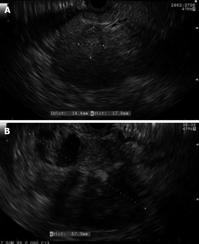
Figure 1 Endoscopic ultrasound image of the heterogenous, nearly isoechoic lesion in the left lobe of the liver (A) and the complex cystic mass in the tail of pancreas (B).
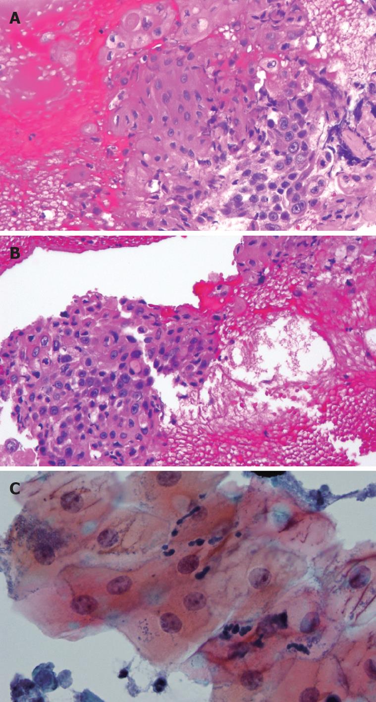
Figure 2 Fine-needle aspiration specimen.
A: Liver mass (HE, × 100); B: Pancreatic mass (HE, × 100). The cell block shows atypical squamous cells consistent with keratinizing squamous cell carcinoma. C: Liver mass (ThinPrep, × 400). Squamous cell contamination from the esophagus as evidenced by the presence of bacteria and fungal organisms.
- Citation: Lai LH, Romagnuolo J, Adams D, Yang J. Primary squamous cell carcinoma of pancreas diagnosed by EUS-FNA: A case report. World J Gastroenterol 2009; 15(34): 4343-4345
- URL: https://www.wjgnet.com/1007-9327/full/v15/i34/4343.htm
- DOI: https://dx.doi.org/10.3748/wjg.15.4343


