Copyright
©2009 The WJG Press and Baishideng.
World J Gastroenterol. Sep 14, 2009; 15(34): 4316-4321
Published online Sep 14, 2009. doi: 10.3748/wjg.15.4316
Published online Sep 14, 2009. doi: 10.3748/wjg.15.4316
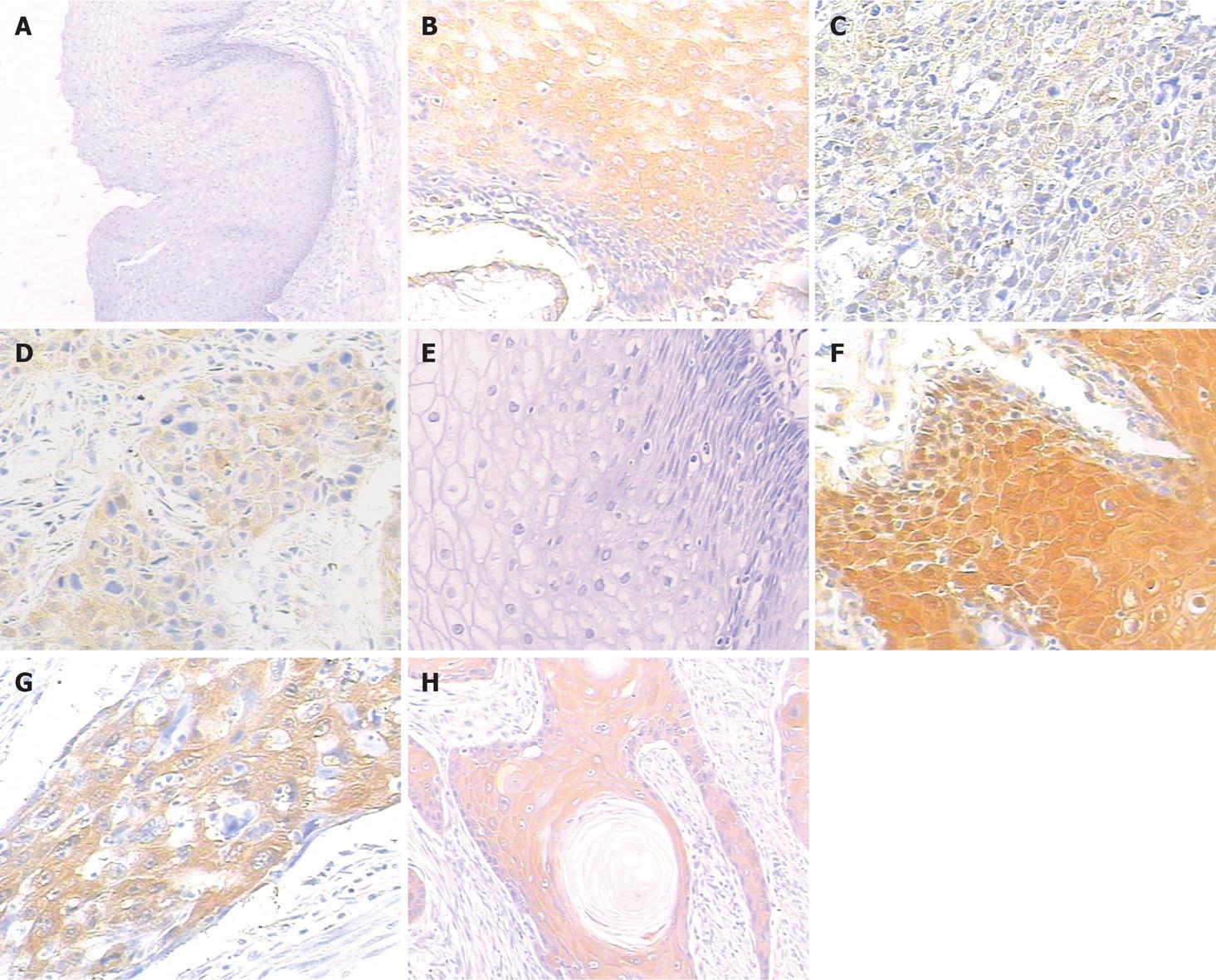
Figure 1 Expression of thymidylate synthase (TS) and glutathione-s-transferase π (GST-π) in normal esophageal mucosa and esophageal squamous cell carcinoma.
A and E: Negative control for TS and GST-π in normal esophageal mucosa, positive staining can not be detected (using TBS as primary antibody) (× 100); B and F: Positive staining was located in the cytoplasm in normal mucosa (× 200); C and G: Poorly-differentiated SCC, note the diffuse strong TS immunostaining in esophageal SCC (× 200), for GST-π, positive staining was shown in the cytoplasm (× 200); D and H: Moderately-differentiated SCC, TS positive staining is in the cytoplasm (× 200); Well-differentiated SCC, diffuse strong GST-π immunostaining can be seen in esophageal SCC (× 200).
- Citation: Huang JX, Li FY, Xiao W, Song ZX, Qian RY, Chen P, Salminen E. Expression of thymidylate synthase and glutathione-s-transferase π in patients with esophageal squamous cell carcinoma. World J Gastroenterol 2009; 15(34): 4316-4321
- URL: https://www.wjgnet.com/1007-9327/full/v15/i34/4316.htm
- DOI: https://dx.doi.org/10.3748/wjg.15.4316


