Copyright
©2009 The WJG Press and Baishideng.
World J Gastroenterol. Aug 28, 2009; 15(32): 4075-4076
Published online Aug 28, 2009. doi: 10.3748/wjg.15.4075
Published online Aug 28, 2009. doi: 10.3748/wjg.15.4075
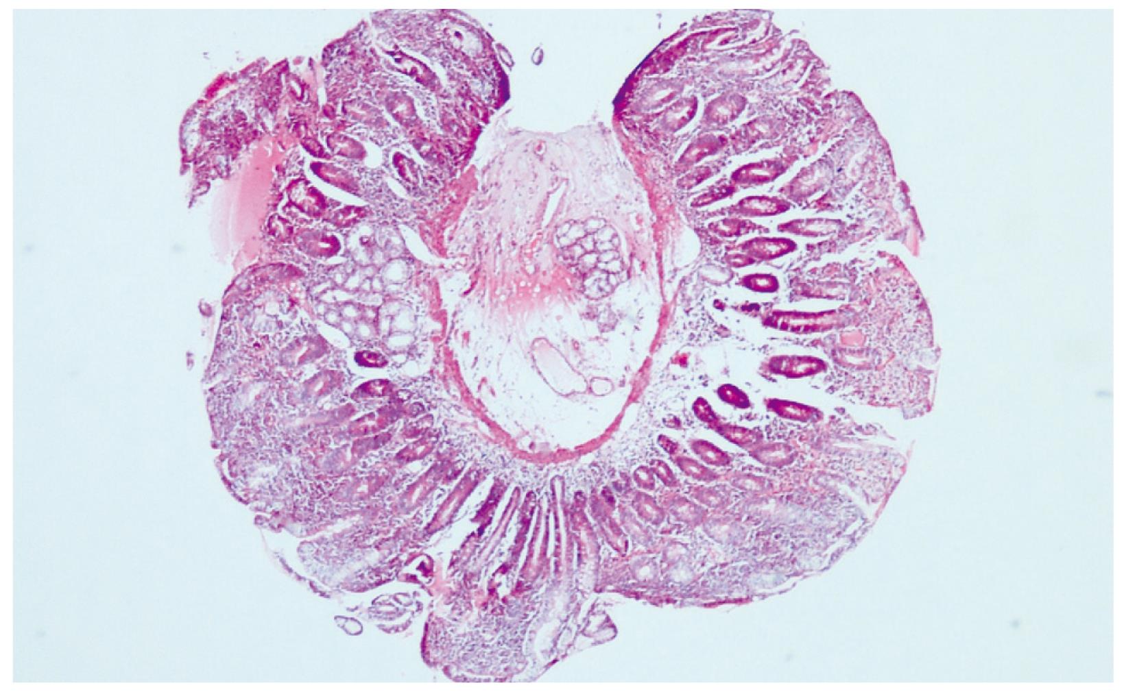
Figure 1 Diffuse villous atrophy in the duodenum (HE, × 10).
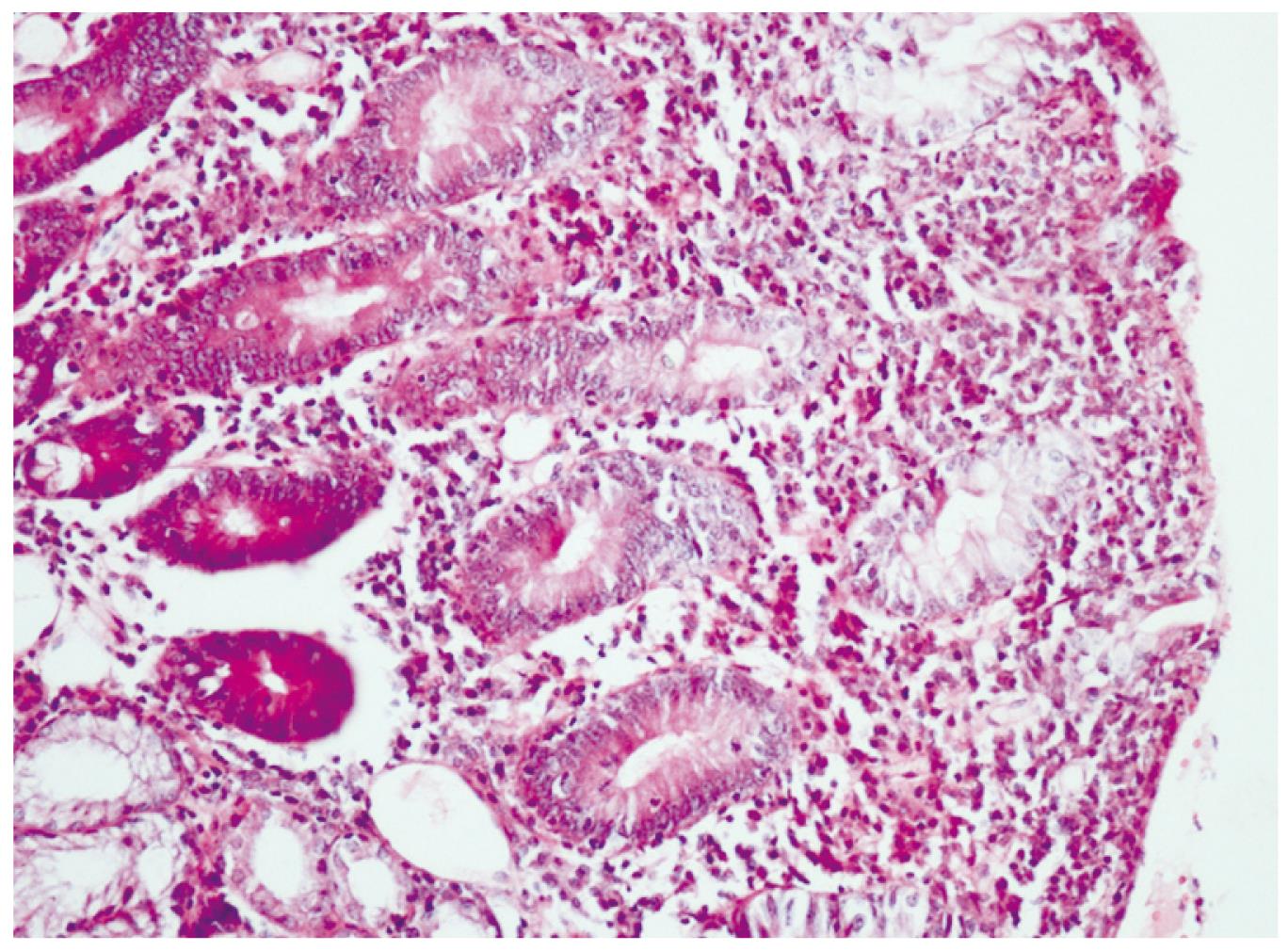
Figure 2 Increased intraepithelial lymphocytes in the duodenum (HE, × 50).
- Citation: Topal F, Akbulut S, Topcu IC, Dolek Y, Yonem O. An adult case of celiac sprue triggered after an ileal resection for perforated Meckel’s diverticulum. World J Gastroenterol 2009; 15(32): 4075-4076
- URL: https://www.wjgnet.com/1007-9327/full/v15/i32/4075.htm
- DOI: https://dx.doi.org/10.3748/wjg.15.4075


