Copyright
©2009 The WJG Press and Baishideng.
World J Gastroenterol. Aug 28, 2009; 15(32): 4009-4015
Published online Aug 28, 2009. doi: 10.3748/wjg.15.4009
Published online Aug 28, 2009. doi: 10.3748/wjg.15.4009
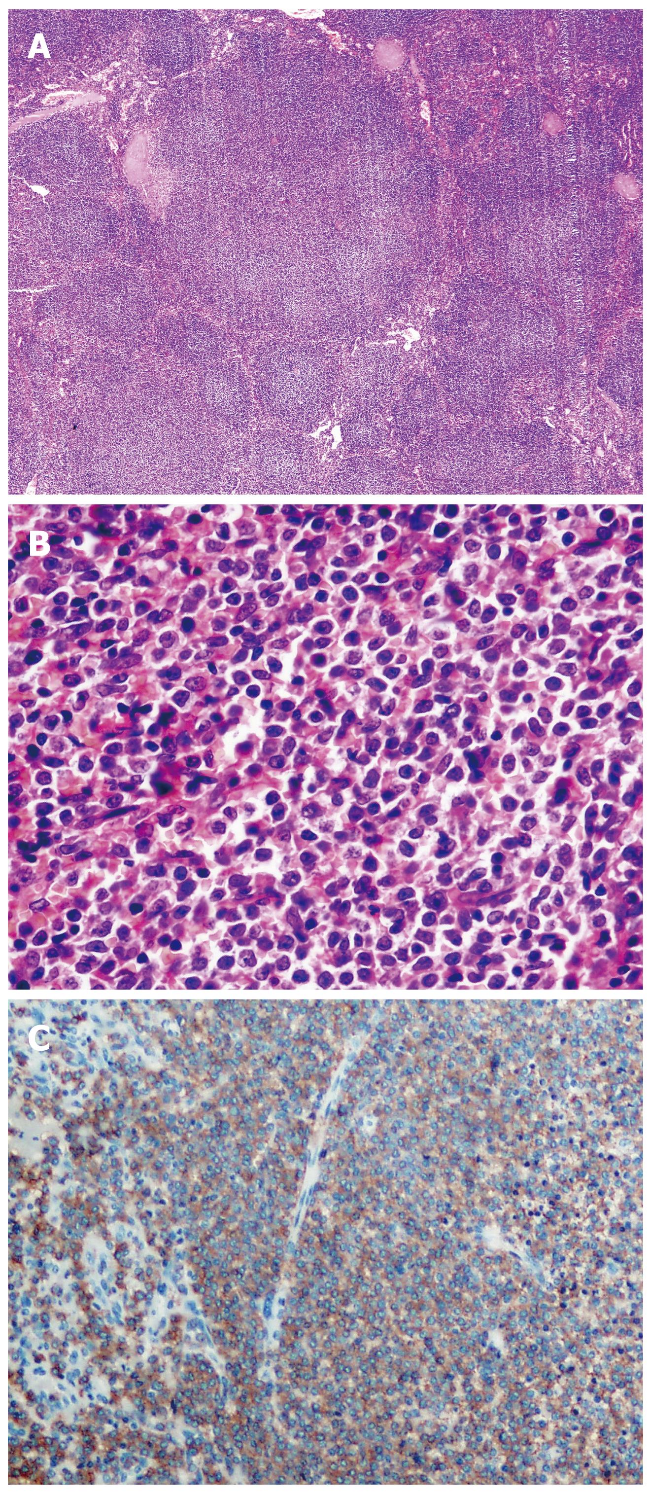
Figure 1 SMZL-splenic tissue.
A: Nodular infiltration (HE, × 40); B: Neoplastic cells with monocytoid morphology (HE, × 400); C: Immunohistochemistry for CD20+, brown staining in lymphoma cells (streptavidin-biotin, × 200).
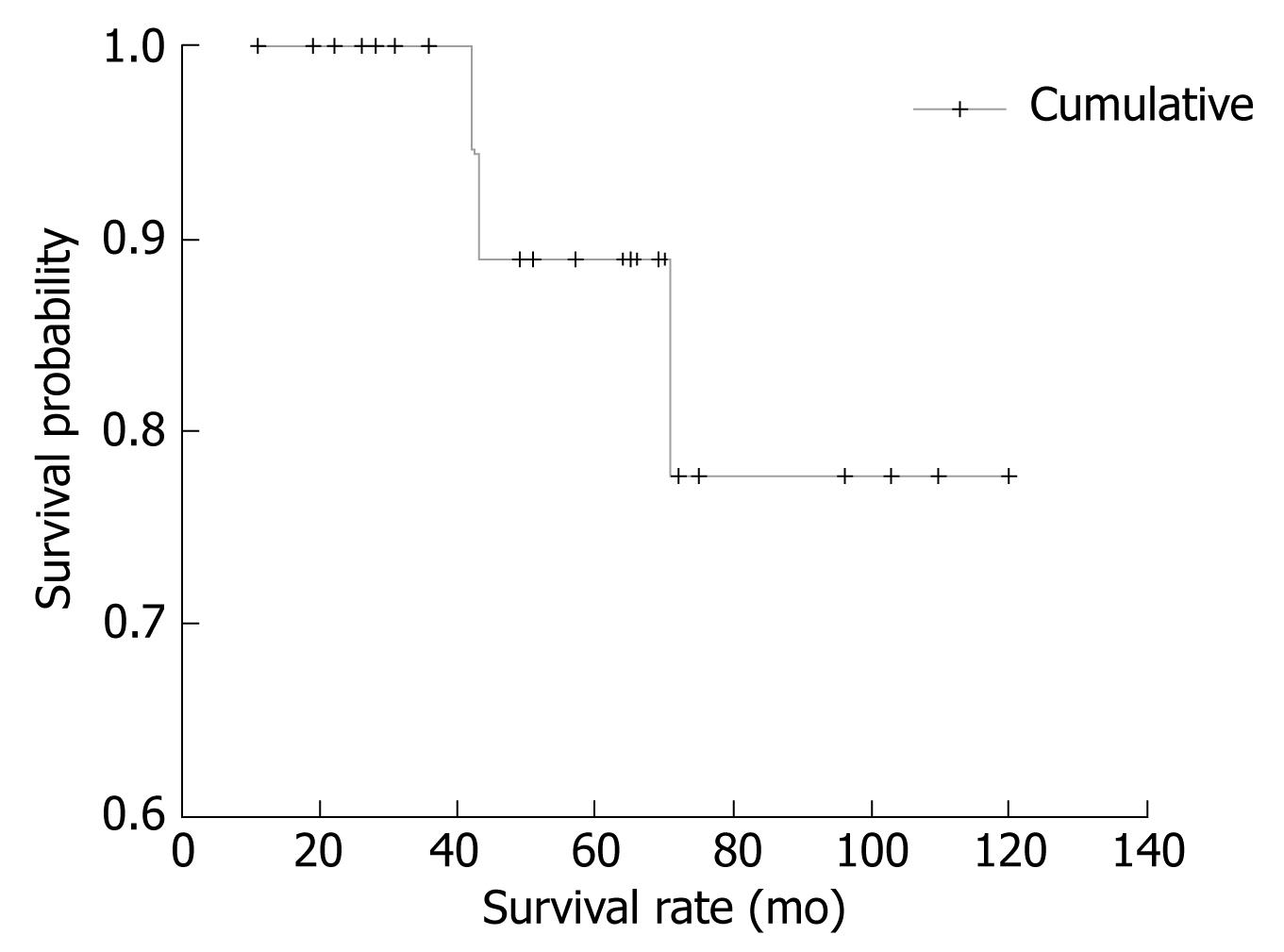
Figure 2 Cumulative survival of patients investigated (with constant incidence of survival after 120 mo).
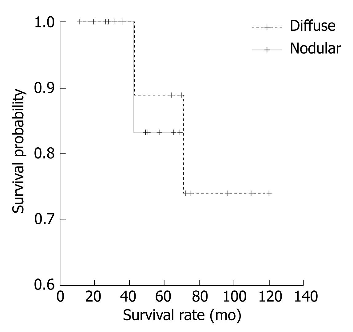
Figure 3 Patients’ survival with different type of spleen infiltration (nodular vs diffuse).
- Citation: Milosevic R, Todorovic M, Balint B, Jevtic M, Krstic M, Ristanovic E, Antonijevic N, Pavlovic M, Perunicic M, Petrovic M, Mihaljevic B. Splenectomy with chemotherapy vs surgery alone as initial treatment for splenic marginal zone lymphoma. World J Gastroenterol 2009; 15(32): 4009-4015
- URL: https://www.wjgnet.com/1007-9327/full/v15/i32/4009.htm
- DOI: https://dx.doi.org/10.3748/wjg.15.4009


