Copyright
©2009 The WJG Press and Baishideng.
World J Gastroenterol. Jan 21, 2009; 15(3): 373-375
Published online Jan 21, 2009. doi: 10.3748/wjg.15.373
Published online Jan 21, 2009. doi: 10.3748/wjg.15.373
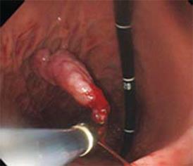
Figure 1 Endoscopy images.
A large lobulated peduncular polyp, about 30 mm in diameter, in the second portion of the duodenum (A and B) and bleeding from the base of the stalk of the polyp (C).
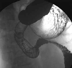
Figure 2 Double contrast radiograph of the duodenum showing an approximately 3-cm peduncular polyp.
Figure 3 Endoscopic polypectomy was performed and the resected specimen was moved into the stomach.
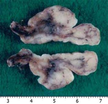
Figure 4 Macroscopic findings of the polypectomy specimen.
The cut surface showed an approximately 3-cm whitish mass.
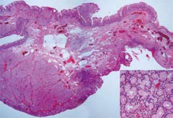
Figure 5 Microscopic findings of the polypectomy specimen.
Low-power view of a cross section showing submucosal proliferation of Brunner’s gland below the duodenal mucosa (HE × 10, × 100).
- Citation: Hirasaki S, Kubo M, Inoue A, Miyake Y, Oshiro H. Pedunculated Brunner’s gland hamartoma of the duodenum causing upper gastrointestinal hemorrhage. World J Gastroenterol 2009; 15(3): 373-375
- URL: https://www.wjgnet.com/1007-9327/full/v15/i3/373.htm
- DOI: https://dx.doi.org/10.3748/wjg.15.373


