Copyright
©2009 The WJG Press and Baishideng.
World J Gastroenterol. Aug 7, 2009; 15(29): 3701-3703
Published online Aug 7, 2009. doi: 10.3748/wjg.15.3701
Published online Aug 7, 2009. doi: 10.3748/wjg.15.3701
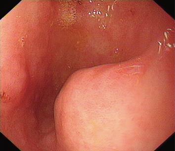
Figure 1 Endoscopy showing a solid tumor mass under the mucosal membrane in the gastric antrum.
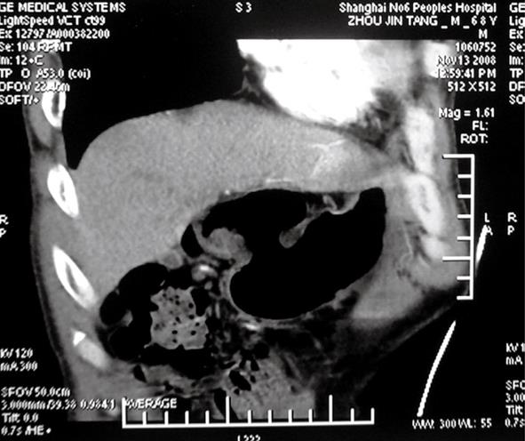
Figure 2 CT reconstruction showing a mass with a diameter of 2 cm in the gastric antrum.
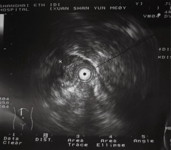
Figure 3 Echogatstroscope revealing low-echogenicity mass under the gastric wall submucosal muscularis propria, with a clear boundary and uneven internal echogenicity.
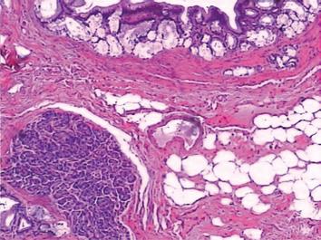
Figure 4 Lobules of pancreatic tissue with ducts located within the smooth muscle of the pylorus (HE, × 100).
- Citation: Yuan Z, Chen J, Zheng Q, Huang XY, Yang Z, Tang J. Heterotopic pancreas in the gastrointestinal tract. World J Gastroenterol 2009; 15(29): 3701-3703
- URL: https://www.wjgnet.com/1007-9327/full/v15/i29/3701.htm
- DOI: https://dx.doi.org/10.3748/wjg.15.3701


