Copyright
©2009 The WJG Press and Baishideng.
World J Gastroenterol. Jul 21, 2009; 15(27): 3448-3450
Published online Jul 21, 2009. doi: 10.3748/wjg.15.3448
Published online Jul 21, 2009. doi: 10.3748/wjg.15.3448
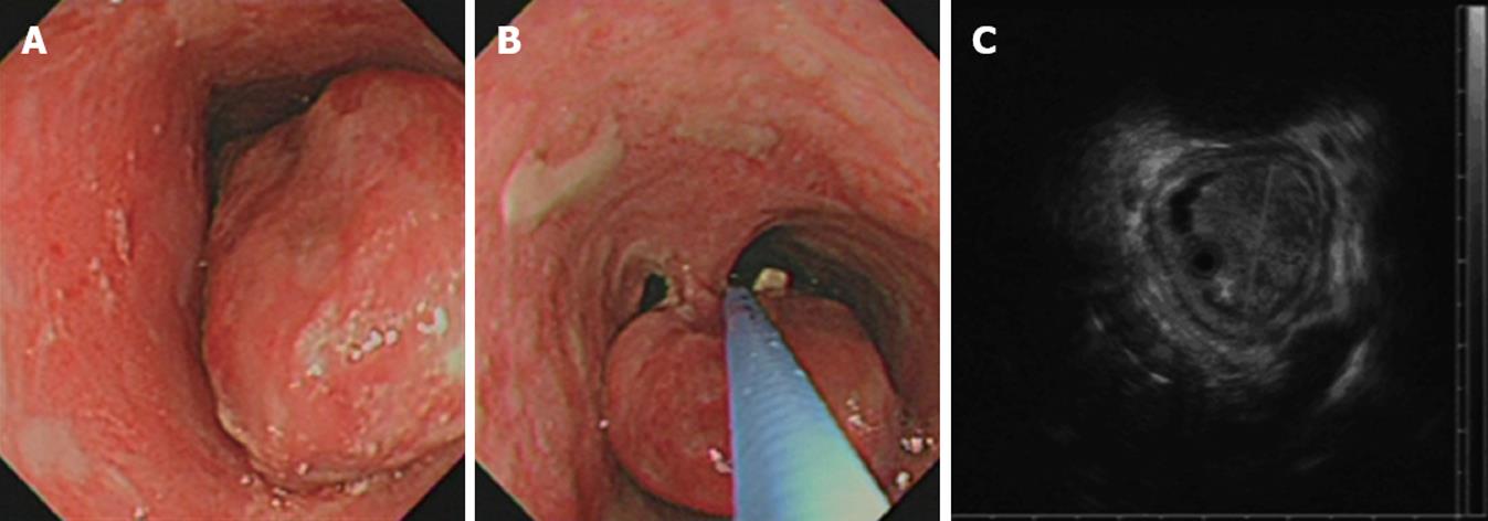
Figure 1 Endoscopic examinations.
A and B: Upper gastrointestinal endoscopy showed a pedunculated polypoid tumor of the esophagus; C: EUS found a 5 cm × 3 cm × 3 cm hypoechoic mass with regular margins confined to mucosal layer only.
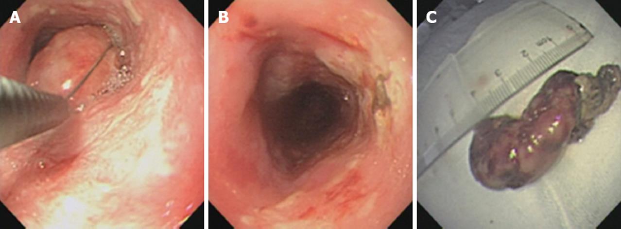
Figure 2 Endoscopic polypectomy.
A: Pre-polypectomy; B: Post-polypectomy; C: Macroscopy of the esophageal carcinosarcoma.
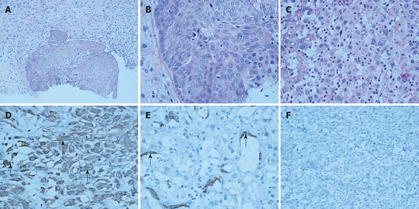
Figure 3 Histological findings.
A: Histology showing a transitional area (HE, × 100); B: A carcinomatous area (HE, × 400); C: A sarcomatous area (HE, × 400); D: Immunohistology showing a sarcomatous area positive for vimentin in cytoplasm (arrows, vimentin, × 400); E: Negative for SMA in cytoplasm and positive for internal control (arrows, SMA, × 400); F: Negative for desmin in cytoplasm (desmin, × 400).
- Citation: Ji F, Xu YM, Xu CF. Endoscopic polypectomy: A promising therapeutic choice for esophageal carcinosarcoma. World J Gastroenterol 2009; 15(27): 3448-3450
- URL: https://www.wjgnet.com/1007-9327/full/v15/i27/3448.htm
- DOI: https://dx.doi.org/10.3748/wjg.15.3448


