Copyright
©2009 The WJG Press and Baishideng.
World J Gastroenterol. Jul 21, 2009; 15(27): 3445-3447
Published online Jul 21, 2009. doi: 10.3748/wjg.15.3445
Published online Jul 21, 2009. doi: 10.3748/wjg.15.3445
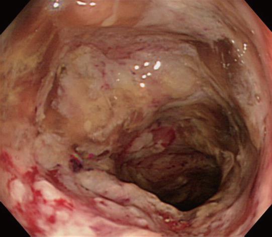
Figure 1 Colonoscopy on admission, showing reddish, friable mucosa and necrotizing ulcers at the transverse colon.
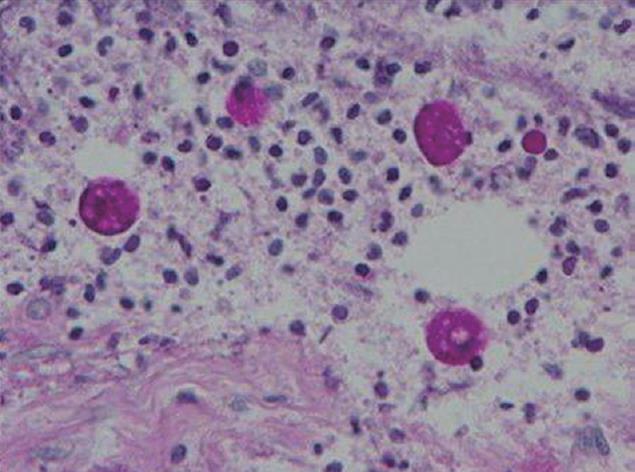
Figure 2 Histological findings of a biopsy specimen, showing Entamoeba that have ingested red blood cells, indicating that they are E.
histolytica.
- Citation: Hanaoka N, Higuchi K, Tanabe S, Sasaki T, Ishido K, Ae T, Koizumi W, Saigenji K. Fulminant amoebic colitis during chemotherapy for advanced gastric cancer. World J Gastroenterol 2009; 15(27): 3445-3447
- URL: https://www.wjgnet.com/1007-9327/full/v15/i27/3445.htm
- DOI: https://dx.doi.org/10.3748/wjg.15.3445


