Copyright
©2009 The WJG Press and Baishideng.
World J Gastroenterol. Jul 21, 2009; 15(27): 3437-3439
Published online Jul 21, 2009. doi: 10.3748/wjg.15.3437
Published online Jul 21, 2009. doi: 10.3748/wjg.15.3437
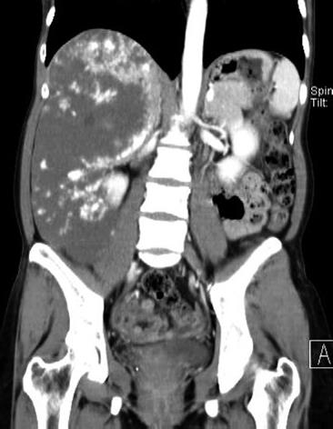
Figure 1 Coronal reformatted CT scan obtained for the portal venous phase shows an ill-defined heterogeneous enhancing mass lesion in the right lobe of the liver.
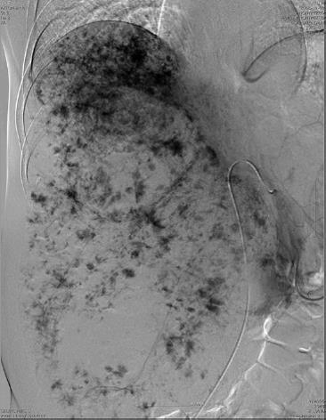
Figure 2 Angiography obtained at the delayed phase, before embolization, shows displacement of vessels and pooling of contrast medium in the right lobe of the liver.
- Citation: Seo HI, Jo HJ, Sim MS, Kim S. Right trisegmentectomy with thoracoabdominal approach after transarterial embolization for giant hepatic hemangioma. World J Gastroenterol 2009; 15(27): 3437-3439
- URL: https://www.wjgnet.com/1007-9327/full/v15/i27/3437.htm
- DOI: https://dx.doi.org/10.3748/wjg.15.3437


