Copyright
©2009 The WJG Press and Baishideng.
World J Gastroenterol. Jun 28, 2009; 15(24): 2975-2979
Published online Jun 28, 2009. doi: 10.3748/wjg.15.2975
Published online Jun 28, 2009. doi: 10.3748/wjg.15.2975
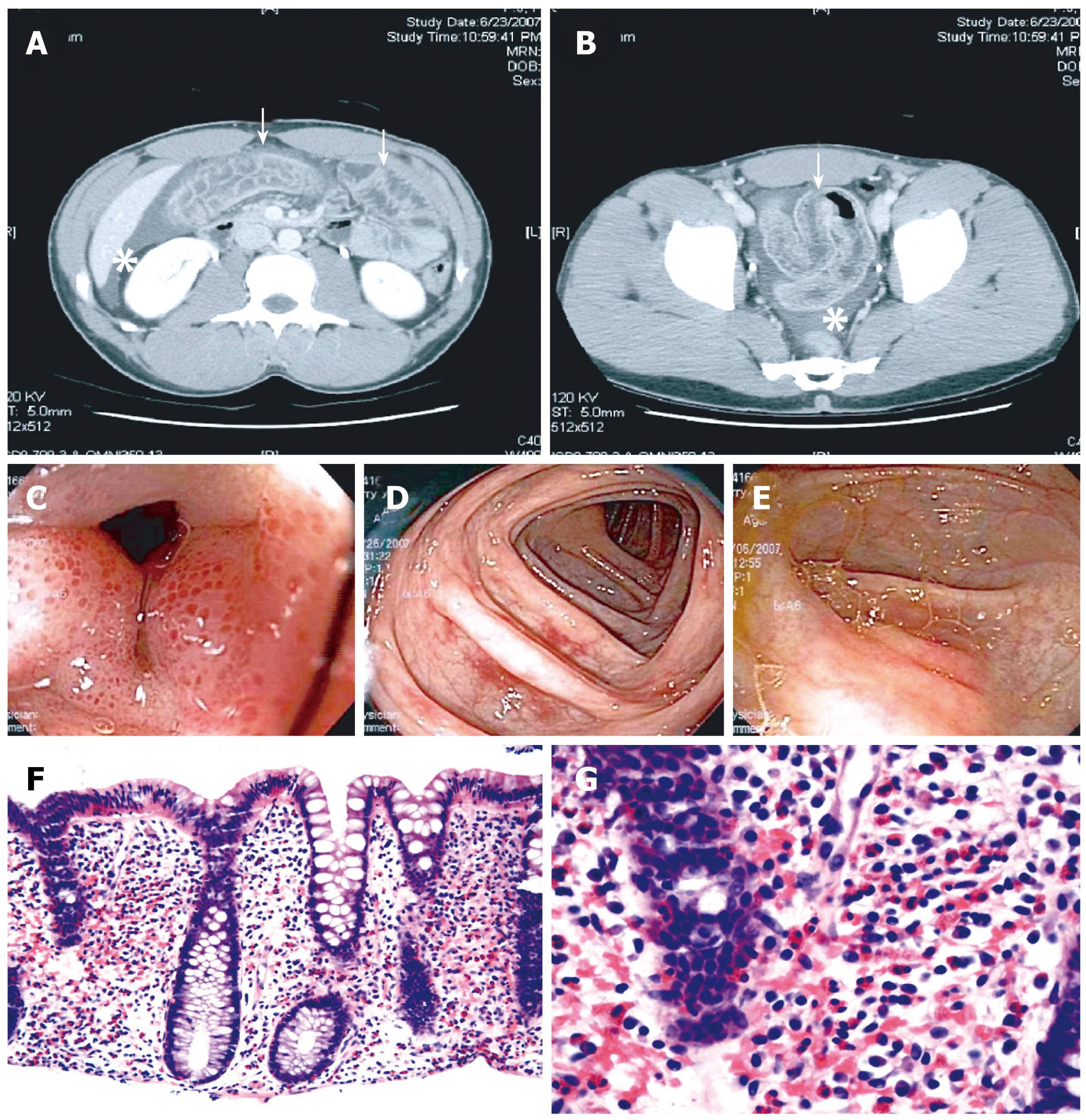
Figure 1 Diagnostic findings in EC.
Representative images from a case of a previously healthy 30-year-old man with recurring episodes of abdominal pain, non-bloody diarrhea, and peripheral eosinophilia; extensive workup confirming EC by exclusion; and excellent response to short-term steroid therapy. A and B: Abdominal CT shows circumferential colon wall thickening (arrows) and moderate ascites (asterisks); C-E: Colonoscopy reveals patchy areas in the colon with mucosal edema and punctate erythema; F and G: Histology indicates markedly increased tissue eosinophilia in all examined segments of the colon. HE stains, magnification 100 × and 400 ×, respectively.
- Citation: Okpara N, Aswad B, Baffy G. Eosinophilic colitis. World J Gastroenterol 2009; 15(24): 2975-2979
- URL: https://www.wjgnet.com/1007-9327/full/v15/i24/2975.htm
- DOI: https://dx.doi.org/10.3748/wjg.15.2975


