Copyright
©2009 The WJG Press and Baishideng.
World J Gastroenterol. Jun 21, 2009; 15(23): 2870-2877
Published online Jun 21, 2009. doi: 10.3748/wjg.15.2870
Published online Jun 21, 2009. doi: 10.3748/wjg.15.2870
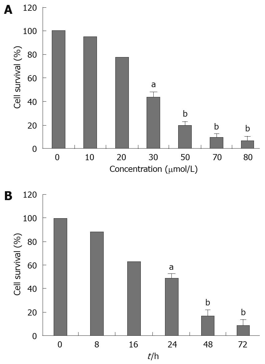
Figure 1 Effect of alisol B acetate on survival of SGC7901 cells.
Cells were incubated in the absence or presence of various concentrations of alisol B acetate for 24 h (A), or at 30 &mgr;mol/L for different incubation times (B). aP < 0.05, bP < 0.01 vs control group (unpaired Student’s t test).
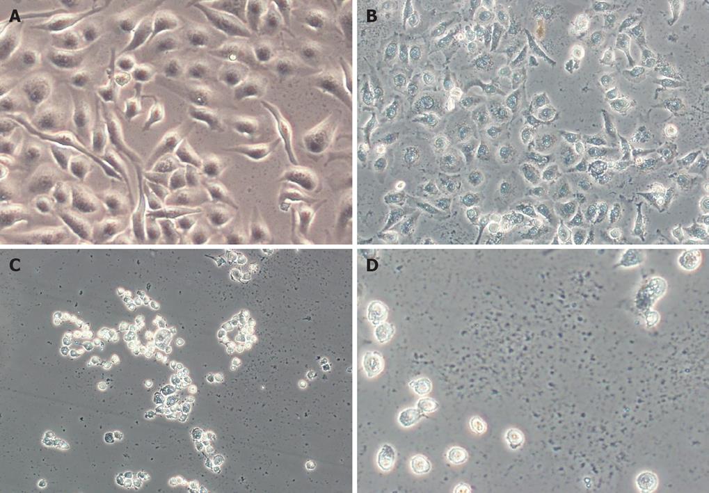
Figure 2 Morphology of SGC7901 cells exposed to alisol B acetate for different concentrations observed under phase-contrast microscope.
A: Controls; B-D: SGC7901 cells were treated with 30, 50 and 70 &mgr;mol/L alisol B acetate for 24 h.
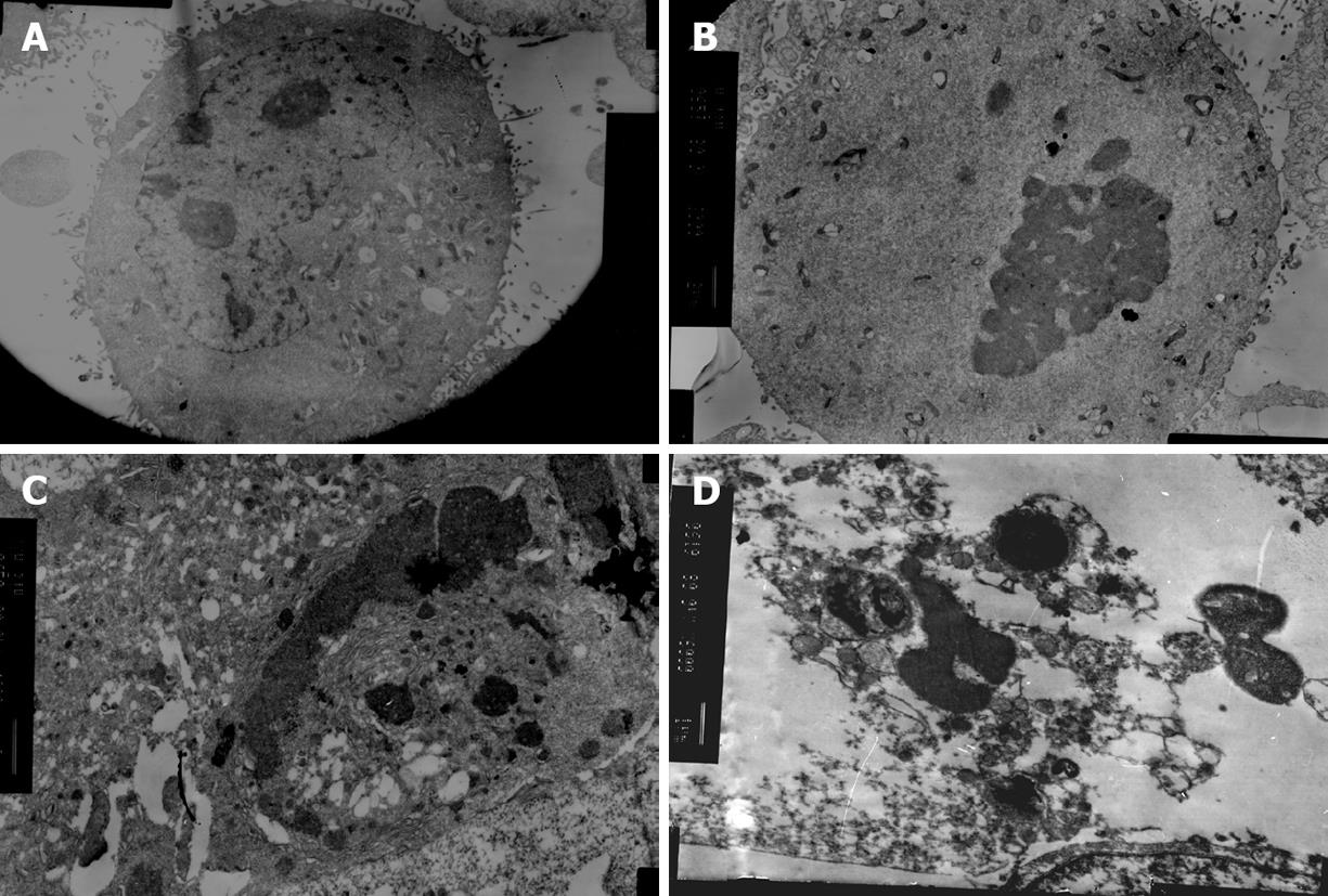
Figure 3 Morphological changes observed under electronic microscope.
A: Control; B-D: Cells were incubated with 30 &mgr;mol/L alisol B acetate for 24, 48 and 72 h.
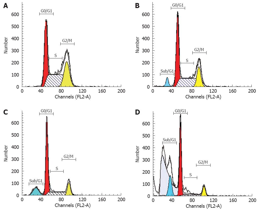
Figure 4 Effect of alisol B acetate on cell cycle distribution of SGC7901 cells.
SGC7901 cells were treated with 0.1% DMSO (A) or with 30 &mgr;mol/L alisol B acetate for 24 h (B), 48 h (C), or 72 h (D). The cells were collected sequentially and stained with PI and analyzed by FCM.
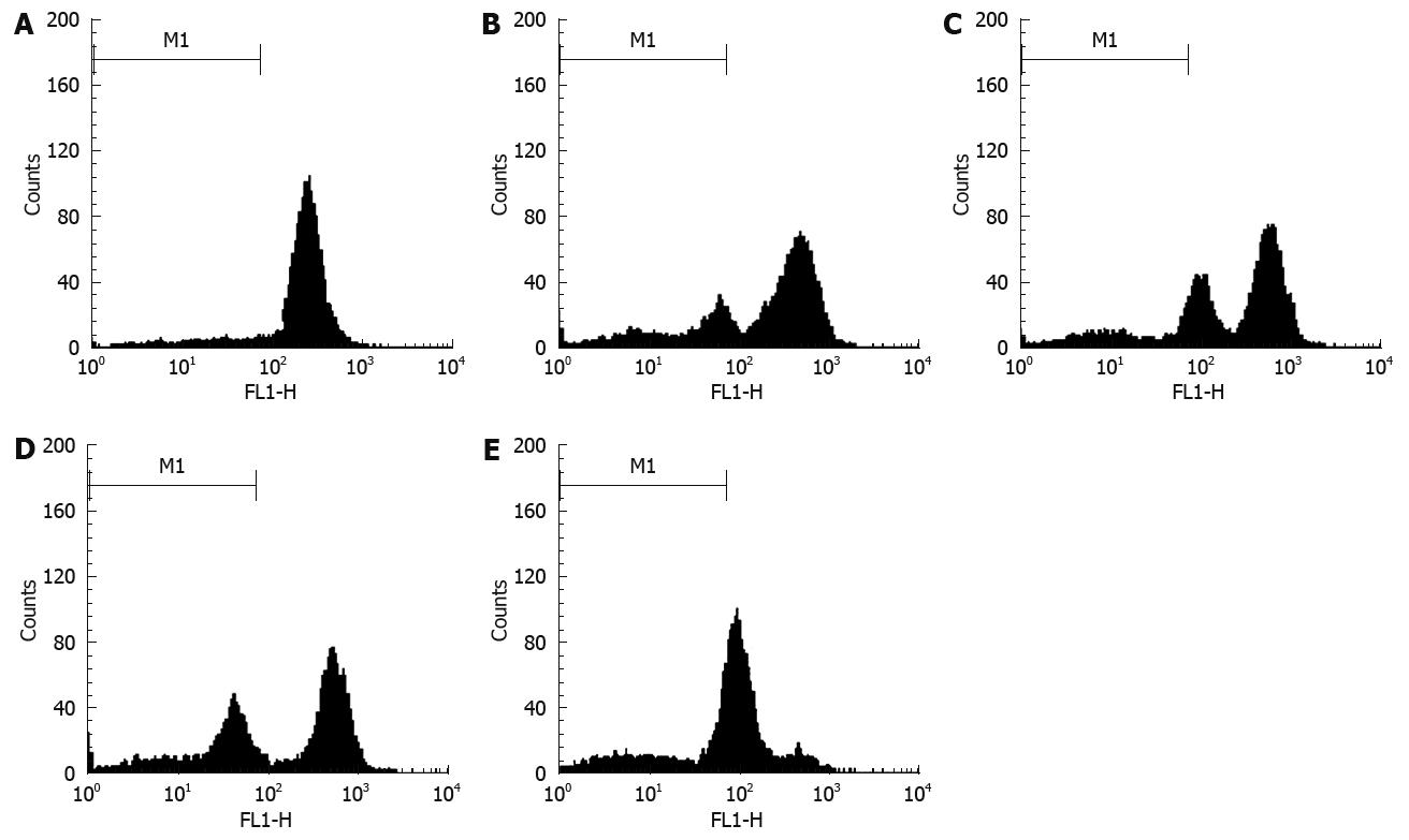
Figure 5 FCM analysis of mitochondrial membrane potential in human SGC7901 cells with 30 &mgr;mol/L alisol B acetate for various time periods.
The zero concentration was defined as control. The percentage of cells stained with Rh-123 was determined by FCM as described in the Materials and Methods. A: Control; B-E: 12, 24, 48 and 72 h.
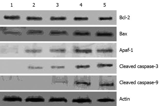
Figure 6 Effect of Alisol B acetate on the expression of Bcl-2, Bax, Apaf-1, cleaved caspase-3 and caspase-9.
SGC7901 cells were treated with 30 &mgr;mol/L alisol B acetate for 12, 24, 48 and 72 h. The cells were collected and lysed. Western blotting analysis was conducted and probed with antibodies to Bcl-2, Bax, Apaf-1, caspase-3 and caspase-9. Lanes (from left to right): Control cells; 12 h; 24 h; 48 h; 72 h.
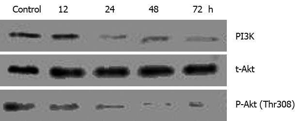
Figure 7 Effect of alisol B acetate on PI3K/Akt activation in SGC7901 cells.
SGC7901 cells were treated with 0.1% DMSO (A) or 30 &mgr;mol/L alisol B acetate for 12, 24, 48 and 72 h. Western blotting analysis for PI3K, t-Akt and P-Akt was performed using specific antibodies with β-actin as a loading control.
-
Citation: Xu YH, Zhao LJ, Li Y. Alisol B acetate induces apoptosis of SGC7901 cells
via mitochondrial and phosphatidylinositol 3-kinases/Akt signaling pathways. World J Gastroenterol 2009; 15(23): 2870-2877 - URL: https://www.wjgnet.com/1007-9327/full/v15/i23/2870.htm
- DOI: https://dx.doi.org/10.3748/wjg.15.2870


