Copyright
©2009 The WJG Press and Baishideng.
World J Gastroenterol. Jun 14, 2009; 15(22): 2739-2747
Published online Jun 14, 2009. doi: 10.3748/wjg.15.2739
Published online Jun 14, 2009. doi: 10.3748/wjg.15.2739
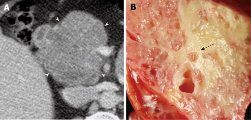
Figure 1 A 64-year-old woman who was incidentally found to have a mass in the head of the pancreas and subsequently underwent a Whipple procedure for removal.
A: Lobulated (arrow heads) microcystic serous cystadenoma (SCA) with characteristic honeycomb appearance is seen on axial MDCT image (1.25 mm); B: Gross pathological specimen of the lesion reveals cluster of microcysts with a sponge pattern. A central scar is appreciated on the pathological image (black arrow), which is subtle and difficult to appreciate on the CT image.
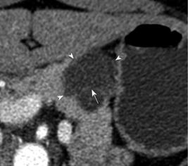
Figure 2 A 46-year-old woman who presented with abdominal pain was found to have a lesion in the body of the pancreas for which a partial pancreatectomy was performed.
Axial MDCT image (1.25 mm) shows a lobulated (arrow heads) macrocystic SCA with septations (white arrow).
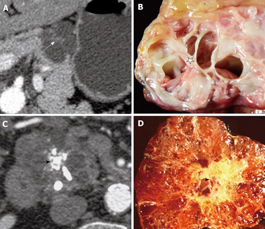
Figure 3 Images from 2 different patients with central scar.
A: Axial MDCT image from a 46-year-old woman with a macrocystic SCA with a central scar (white arrow); B: Gross pathological specimen of the same patient also reveals the macrocystic pattern with the septa converging on a central scar (star); C: Axial MDCT image from a 66-year-old male who had a history of chronic pancreatitis, reveals a large lobulated microcystic lesion with central stellate calcification (black arrow). A Whipple procedure was done to remove a 4 cm mass from the head of the pancreas. Corresponding gross pathological image of the microcystic SCA with calcification (black arrow) in the central scar (D).
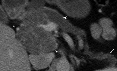
Figure 4 Axial MDCT image from a 64-year-old woman who had a large cyst in the pancreas detected incidentally.
The patient underwent a Whipple procedure for removal of the large microcystic SCA (arrow heads) involving the head and uncinate process of the pancreas. The SCA caused upstream pancreatic duct dilatation (white arrow) and atrophy of the pancreatic body and tail (small white arrow).
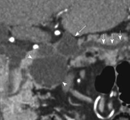
Figure 5 Coronal reformatted MDCT image from an asymptomatic 90-year-old woman, who had a 5 cm lesion in the head of the pancreas which was removed by Whipple procedure.
The image shows an oligocystic SCA (arrow heads) in the head of pancreas which was mistaken for a mucinous lesion due to an associated side branch IPMN adjacent to it (white arrow). The two lesions were interpreted as a single multiloculated side branch mucinous lesion. There is mild upstream dilatation of the visualized pancreatic duct (small white arrows).
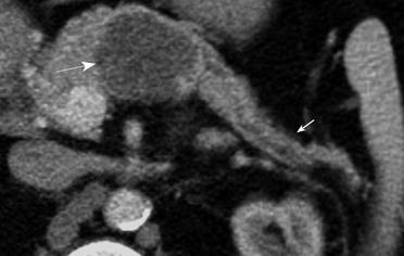
Figure 6 Axial MDCT image from a 64-year-old woman with an incidentally discovered cystic lesion in the body of the pancreas.
A large serous cystadenoma (arrow) present in the body of pancreas displays microcystic morphology and fine lobulations with a central scar. Atrophy of the pancreatic parenchyma distal to the site of the lesion is also present (small arrow).
- Citation: Shah AA, Sainani NI, Ramesh AK, Shah ZK, Deshpande V, Hahn PF, Sahani DV. Predictive value of multi-detector computed tomography for accurate diagnosis of serous cystadenoma: Radiologic-pathologic correlation. World J Gastroenterol 2009; 15(22): 2739-2747
- URL: https://www.wjgnet.com/1007-9327/full/v15/i22/2739.htm
- DOI: https://dx.doi.org/10.3748/wjg.15.2739


