Copyright
©2009 The WJG Press and Baishideng.
World J Gastroenterol. May 21, 2009; 15(19): 2357-2360
Published online May 21, 2009. doi: 10.3748/wjg.15.2357
Published online May 21, 2009. doi: 10.3748/wjg.15.2357
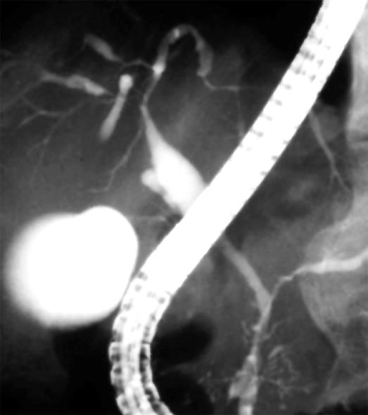
Figure 1 Endoscopic retrograde cholangiography of a patient with autoimmune pancreatitis showing a relatively long stricture of the hepatic hilar bile duct.
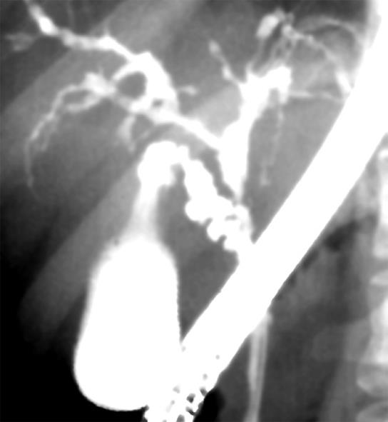
Figure 2 Endoscopic retrograde cholangiography of a patient with primary sclerosing cholangitis showing beaded and pruned-tree appearance.
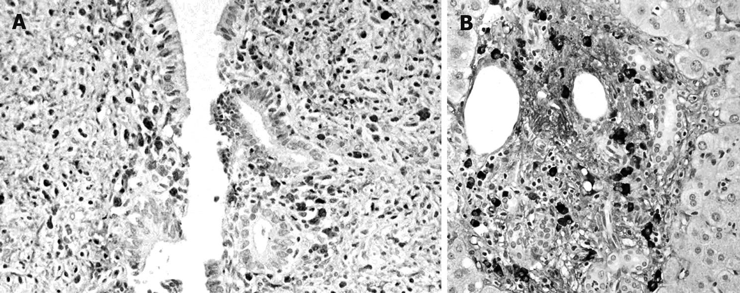
Figure 3 IgG4-immunostaining of the bile duct (A) and liver (B) of a patient with autoimmune pancreatitis.
Dense infiltration of IgG4-positive plasma cells was detected in the bile duct wall (A) and the periportal area of the liver (B).
- Citation: Kamisawa T, Takuma K, Anjiki H, Egawa N, Kurata M, Honda G, Tsuruta K. Sclerosing cholangitis associated with autoimmune pancreatitis differs from primary sclerosing cholangitis. World J Gastroenterol 2009; 15(19): 2357-2360
- URL: https://www.wjgnet.com/1007-9327/full/v15/i19/2357.htm
- DOI: https://dx.doi.org/10.3748/wjg.15.2357


