Copyright
©2009 The WJG Press and Baishideng.
World J Gastroenterol. May 14, 2009; 15(18): 2296-2299
Published online May 14, 2009. doi: 10.3748/wjg.15.2296
Published online May 14, 2009. doi: 10.3748/wjg.15.2296
Figure 1 Contrast-enhanced CT.
Heterogeneous, not diffuse, hypervascular nodule in the early phase (A) (arrow), and a low-density area in the late phase (B) (arrow).
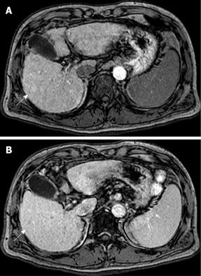
Figure 2 Contrast-enhanced MRI.
A heterogeneous hypervascular nodule in the early phase (A) (arrow), and washout in the late phase (B) (arrow).
Figure 3 Contrast-enhanced US.
Heterogeneous hypervascularity in the early phase (A) (arrow), and defect in the Kupffer phase (B) (arrow).
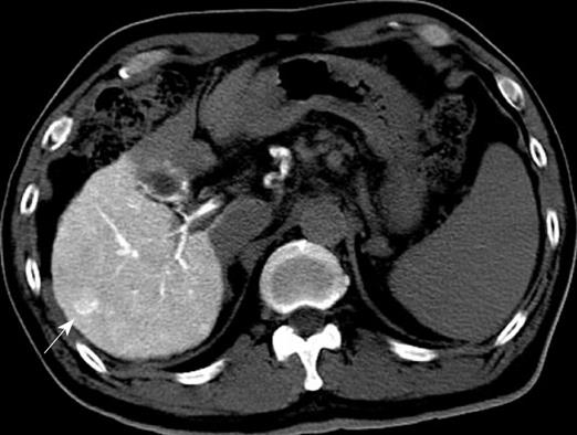
Figure 4 Heterogeneous hyperattenuation at CTA (arrow).
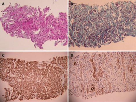
Figure 5 US-guided biopsy.
Moderately-differentiated HCC characterized by typical cytological and structural atypia with dense fibrosis (HE stain) (A), (Mallory-Azan stain) (B). Positive for Hp1 (C) and α-SMA (D).
- Citation: Kim SR, Imoto S, Nakajima T, Ando K, Mita K, Fukuda K, Nishikawa R, Koma YI, Matsuoka T, Kudo M, Hayashi Y. Scirrhous hepatocellular carcinoma displaying atypical findings on imaging studies. World J Gastroenterol 2009; 15(18): 2296-2299
- URL: https://www.wjgnet.com/1007-9327/full/v15/i18/2296.htm
- DOI: https://dx.doi.org/10.3748/wjg.15.2296


