Copyright
©2009 The WJG Press and Baishideng.
World J Gastroenterol. May 14, 2009; 15(18): 2283-2286
Published online May 14, 2009. doi: 10.3748/wjg.15.2283
Published online May 14, 2009. doi: 10.3748/wjg.15.2283
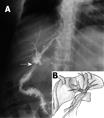
Figure 1 T-tube cholangiography.
A: Postero-anterior T-tube cholangiogram showing biliary tract mal-repair (arrowhead indicates the T-tube, arrow indicates the tube drain); B: Schematic diagram of the cholangiogram.
Figure 2 T-tube cholangiography.
A: Postero-anterior view of the T-tube cholangiogram, which was done in our hospital, after the definitive surgical repair of the biliary tract injury (arrow: two intra-bilio-jejunal stents); B: Schematic diagram of the cholangiogram.
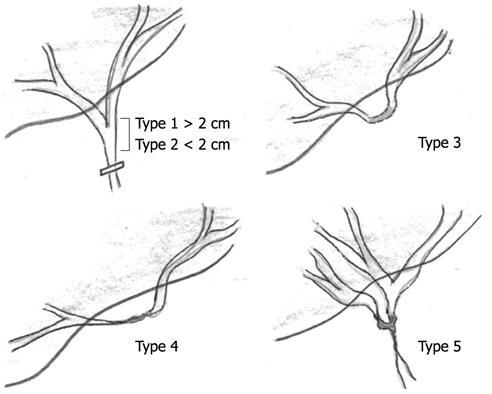
Figure 3 Bismuth classification of biliary injuries.
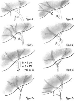
Figure 4 Strasberg classification of biliary injuries.
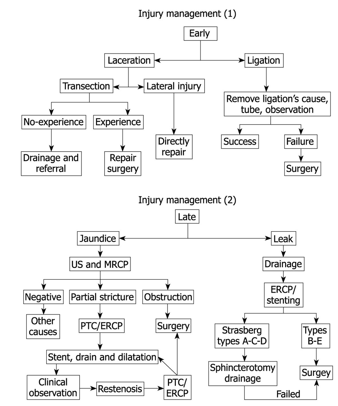
Figure 5 Our suggested flow chart for the management of early and late presentations of biliary injuries.
PTC: Percutaneous transhepatic cholangiography; ERCP: Endoscopic retrograde cholangiopancreatography.
- Citation: Aldumour A, Aseni P, Alkofahi M, Lamperti L, Aldumour E, Girotti P, Carlis LGD. Repair of a mal-repaired biliary injury: A case report. World J Gastroenterol 2009; 15(18): 2283-2286
- URL: https://www.wjgnet.com/1007-9327/full/v15/i18/2283.htm
- DOI: https://dx.doi.org/10.3748/wjg.15.2283


