Copyright
©2009 The WJG Press and Baishideng.
World J Gastroenterol. Apr 7, 2009; 15(13): 1630-1635
Published online Apr 7, 2009. doi: 10.3748/wjg.15.1630
Published online Apr 7, 2009. doi: 10.3748/wjg.15.1630
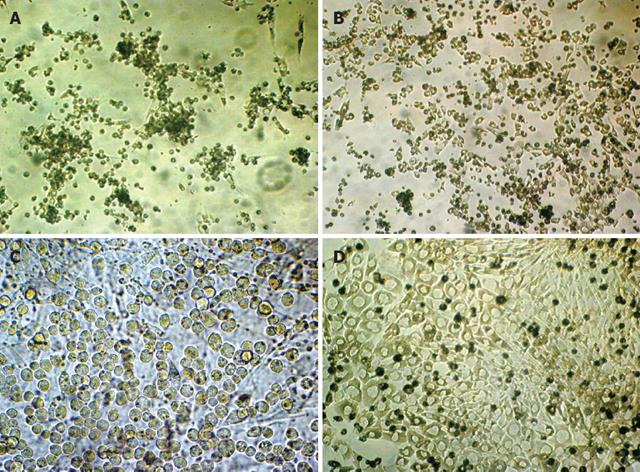
Figure 1 Morphological evidence of BDLSC diff-erentiation.
A: BDLSC clone selected from bone marrow cells, phase-contrast microscope (200 ×); B: Proliferation of liver stem cell clone-phase-contrast microscope (200 ×); C: Differentiation-prohibited passage stem cells, phase-contrast microscope (400 ×); D: Hepatocyte-like cells after differentiation, phase-contrast microscope (400 ×).
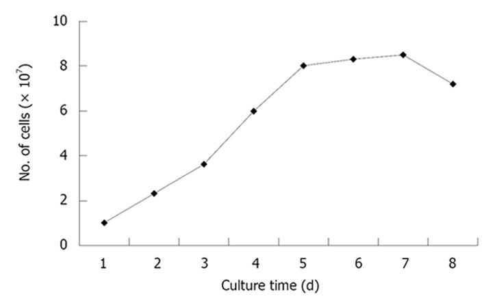
Figure 2 Cell growth curve of passaged BDLSC.
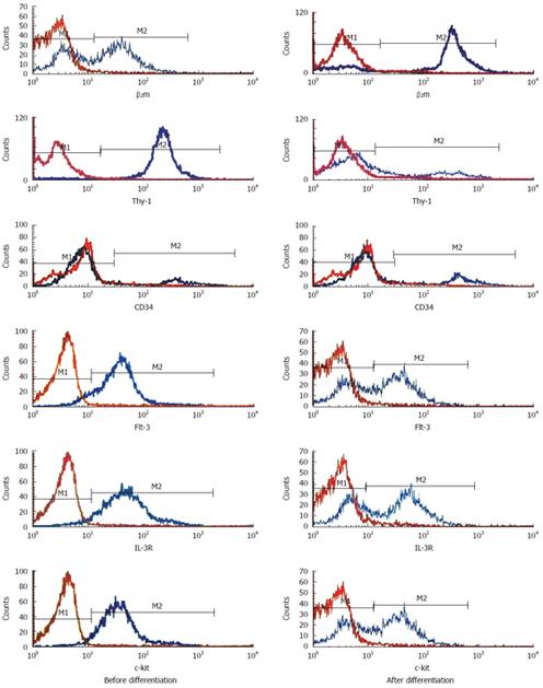
Figure 3 Differences of stem cell markers before and after differentiation by flow cytometry.
Red curves: Negative control, M1: Negative part, M2: Positive part.
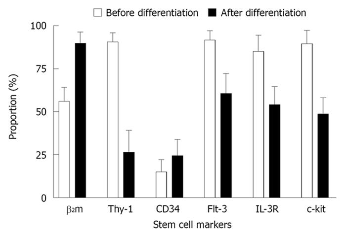
Figure 4 Expression of stem cell markers before and after differentiation.
- Citation: Cai YF, Chen JS, Su SY, Zhen ZJ, Chen HW. Passage of bone-marrow-derived liver stem cells in a proliferating culture system. World J Gastroenterol 2009; 15(13): 1630-1635
- URL: https://www.wjgnet.com/1007-9327/full/v15/i13/1630.htm
- DOI: https://dx.doi.org/10.3748/wjg.15.1630


