Copyright
©2009 The WJG Press and Baishideng.
World J Gastroenterol. Mar 28, 2009; 15(12): 1499-1505
Published online Mar 28, 2009. doi: 10.3748/wjg.15.1499
Published online Mar 28, 2009. doi: 10.3748/wjg.15.1499
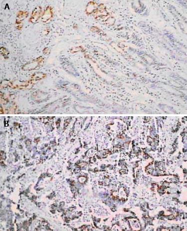
Figure 1 Immunohistochemical staining.
A: PRL-3 is negative or weak in adjacent (3 cm away from the tumor) normal gastric epithelial mucosa (× 40); B: In positive cases, PRL-3 expression in cancer cell cytoplasms is strong (× 200).
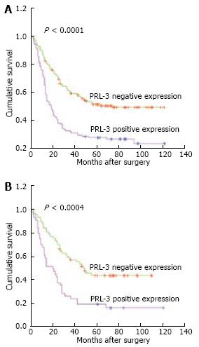
Figure 2 Overall survival curve.
A: Entire cohort of 293 patients; B: Patients with stage III. Significant differences were observed between the two groups with PRL-3 negative and positive expression.
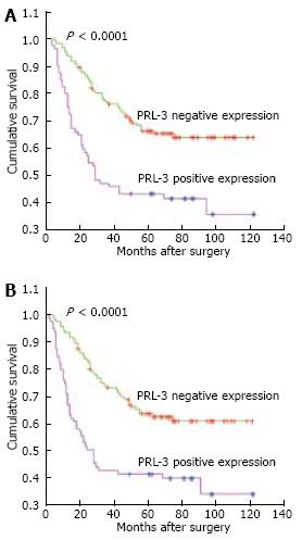
Figure 3 Patients who underwent curative surgery.
A: Overall survival; B: Disease-free survival. Significant differences were observed between the PRL-3 negative and positive groups.
- Citation: Dai N, Lu AP, Shou CC, Li JY. Expression of phosphatase regenerating liver 3 is an independent prognostic indicator for gastric cancer. World J Gastroenterol 2009; 15(12): 1499-1505
- URL: https://www.wjgnet.com/1007-9327/full/v15/i12/1499.htm
- DOI: https://dx.doi.org/10.3748/wjg.15.1499


