Copyright
©2009 The WJG Press and Baishideng.
World J Gastroenterol. Mar 28, 2009; 15(12): 1452-1458
Published online Mar 28, 2009. doi: 10.3748/wjg.15.1452
Published online Mar 28, 2009. doi: 10.3748/wjg.15.1452
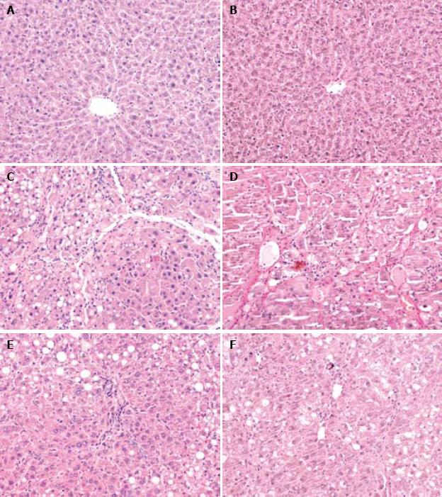
Figure 1 Pathological changes.
Light microscopy of liver tissue sections showing normal liver lobular architecture with central veins in the normal control group (HE staining, × 200) (A), no collagen deposition in liver of normal control group (VG staining, × 200) (B), degenerated and necrotic liver cells associated with inflammatory cells in model group (HE staining, × 200) (C), formation of fibrotic septa in model group (VG staining, × 200) (D), and pathological change in liver of CCl4 + melatonin (10 mg/kg) group was rather milder compared with the model group (HE staining, × 200; VG staining, × 200) (E, F).
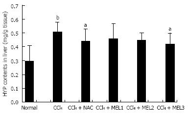
Figure 2 Effect of melatonin on hydroxyproline content in liver of rats with fibrosis fibrotic induced by CCl4.
aP < 0.05 vs model control group; bP < 0.01 vs normal control group.
- Citation: Hong RT, Xu JM, Mei Q. Melatonin ameliorates experimental hepatic fibrosis induced by carbon tetrachloride in rats. World J Gastroenterol 2009; 15(12): 1452-1458
- URL: https://www.wjgnet.com/1007-9327/full/v15/i12/1452.htm
- DOI: https://dx.doi.org/10.3748/wjg.15.1452


