Copyright
©2009 The WJG Press and Baishideng.
World J Gastroenterol. Mar 21, 2009; 15(11): 1388-1390
Published online Mar 21, 2009. doi: 10.3748/wjg.15.1388
Published online Mar 21, 2009. doi: 10.3748/wjg.15.1388
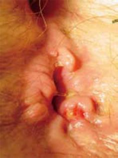
Figure 1 The lesion as it appeared macroscopically at the time of diagnosis.
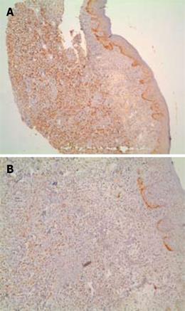
Figure 2 Positive immunohistochemical staining in anal lesion.
A: ERs (× 4); B: PRs ( × 10).
- Citation: Puglisi M, Varaldo E, Assalino M, Ansaldo G, Torre G, Borgonovo G. Anal metastasis from recurrent breast lobular carcinoma: A case report. World J Gastroenterol 2009; 15(11): 1388-1390
- URL: https://www.wjgnet.com/1007-9327/full/v15/i11/1388.htm
- DOI: https://dx.doi.org/10.3748/wjg.15.1388


