Copyright
©2008 The WJG Press and Baishideng.
World J Gastroenterol. Mar 7, 2008; 14(9): 1459-1462
Published online Mar 7, 2008. doi: 10.3748/wjg.14.1459
Published online Mar 7, 2008. doi: 10.3748/wjg.14.1459
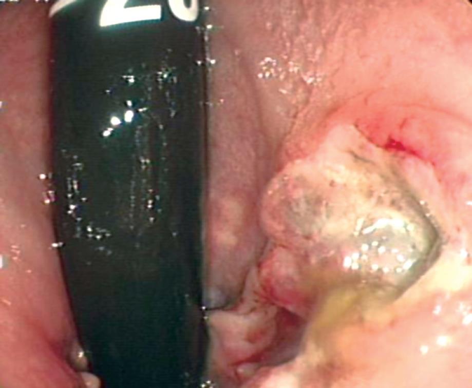
Figure 1 On colonoscopy, an ulcerated fungating mass was detected 3 cm from the anal verge.
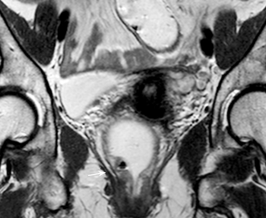
Figure 2 Magnetic resonance imaging shows a T1-weighted of low signal area (arrow) growing in the anterior portion of the anorectal junction.
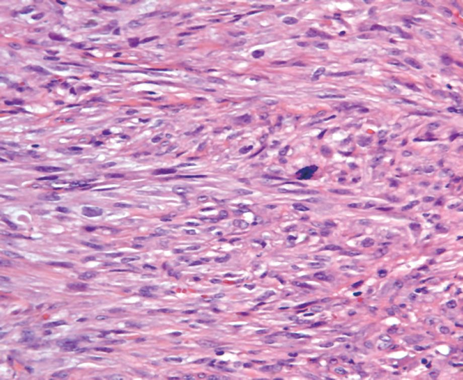
Figure 3 Histological section shows a typical malignant fibrous histiocytoma featuring spindle cells arranged in a storiform pattern (HE, × 400).
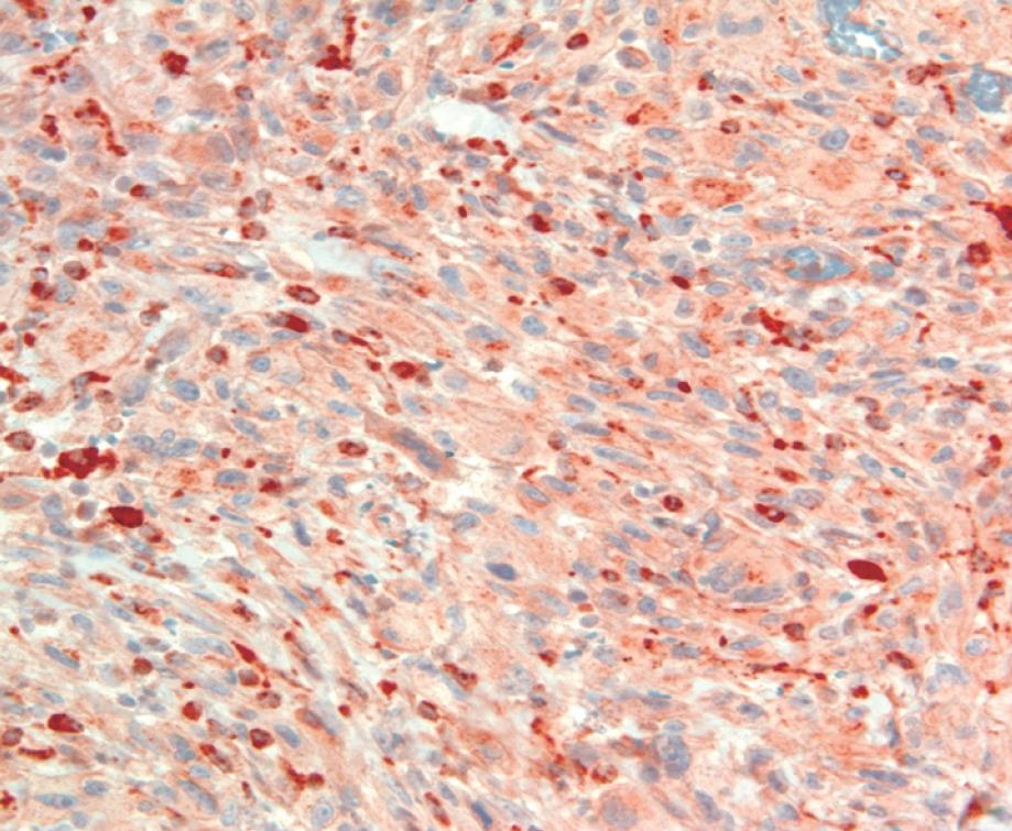
Figure 4 Tumor cells show positive immunostaining for CD68 (× 400).
- Citation: Kim BG, Chang IT, Park JS, Choi YS, Kim GH, Park ES, Choi CH. Transanal excision of a malignant fibrous histiocytoma of anal canal: A case report and literature review. World J Gastroenterol 2008; 14(9): 1459-1462
- URL: https://www.wjgnet.com/1007-9327/full/v14/i9/1459.htm
- DOI: https://dx.doi.org/10.3748/wjg.14.1459


