Copyright
©2008 The WJG Press and Baishideng.
World J Gastroenterol. Mar 7, 2008; 14(9): 1453-1455
Published online Mar 7, 2008. doi: 10.3748/wjg.14.1453
Published online Mar 7, 2008. doi: 10.3748/wjg.14.1453
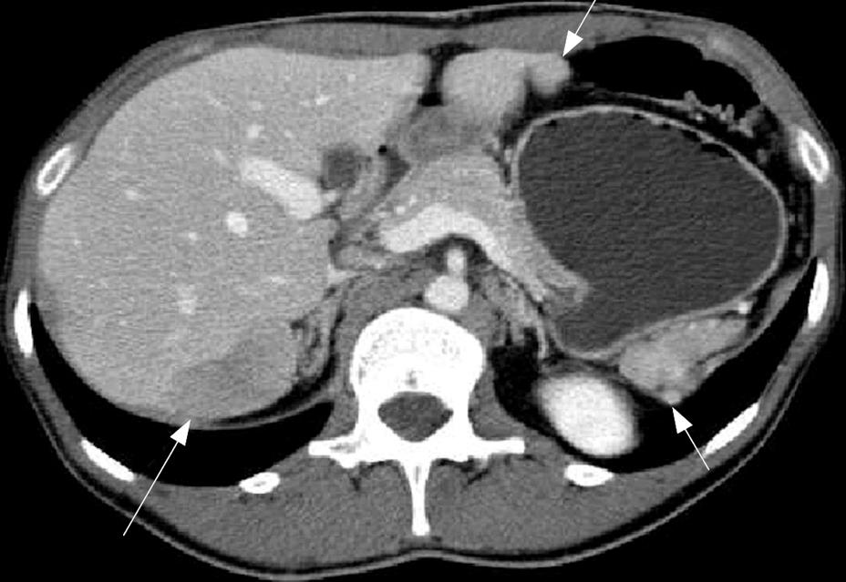
Figure 1 Contrast-enhanced helical CT obtained during the portal phase of acquisition, showing a hypodense 3 cm lesion along the posterior surface of the seventh segment of the right lobe of the liver (long white arrow), and two similar nodular lesions medially to the left lobe of the liver and adjacent to the upper pole of the left kidney and the pancreatic tail (small white arrows).
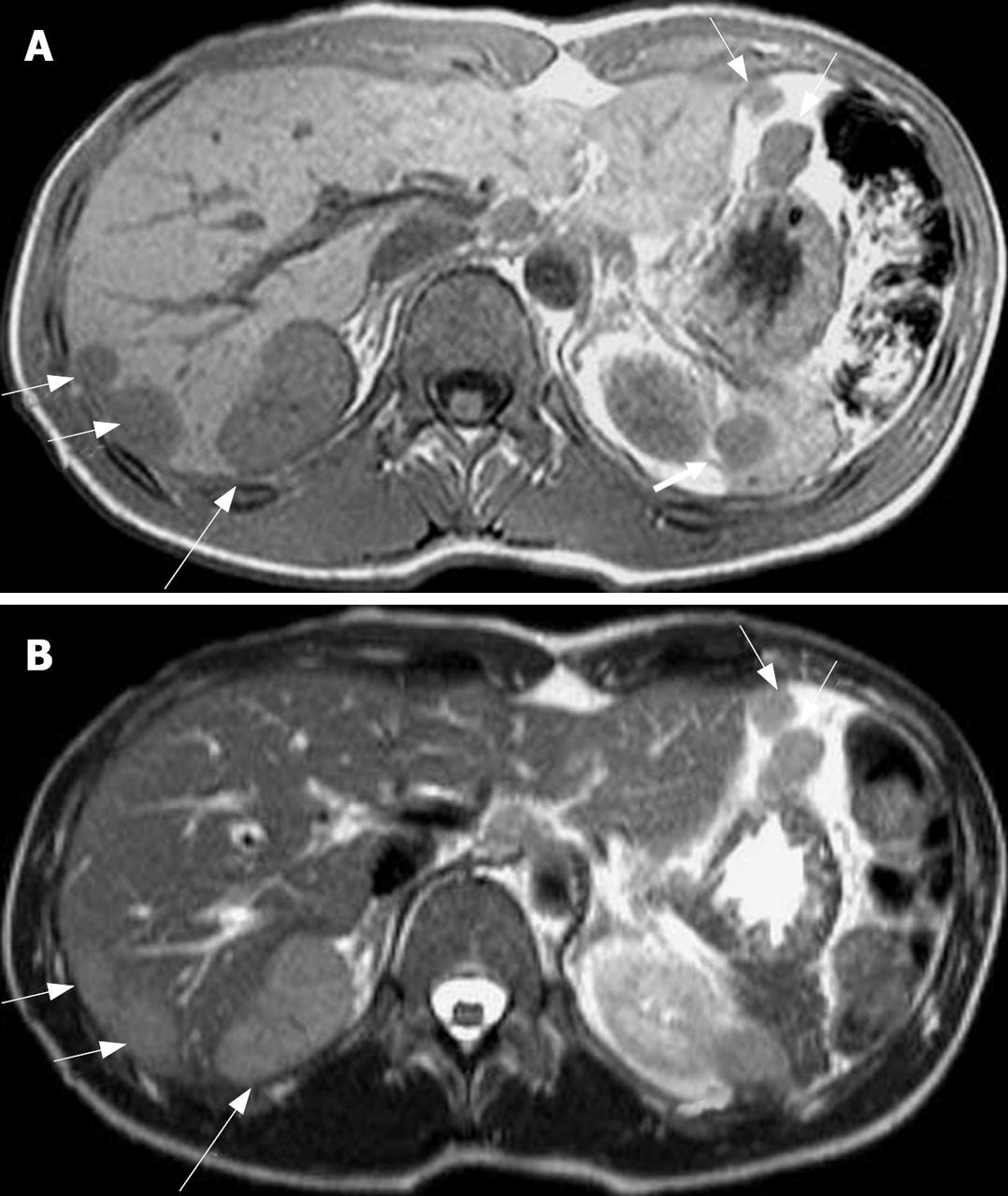
Figure 2 Different image showing a hypointense 3 cm lesion along the posterior surface of the seventh segment of the right lobe of the liver (long white arrow) and 5 additional lesions in the sub-capsular portion of the seventh segment of the liver, medially to the left lobe of the liver and adjacent to the upper pole of the left kidney and the pancreatic tail (small white arrows).
A: Unenhanced T1-weighted (TR: 218, TE: 4.6 ms) axial MRI scan; B: T2-weighted (TR: 417, TE: 80 ms) axial image.
- Citation: Imbriaco M, Camera L, Manciuria A, Salvatore M. A case of multiple intra-abdominal splenosis with computed tomography and magnetic resonance imaging correlative findings. World J Gastroenterol 2008; 14(9): 1453-1455
- URL: https://www.wjgnet.com/1007-9327/full/v14/i9/1453.htm
- DOI: https://dx.doi.org/10.3748/wjg.14.1453


