Copyright
©2008 The WJG Press and Baishideng.
World J Gastroenterol. Mar 7, 2008; 14(9): 1437-1443
Published online Mar 7, 2008. doi: 10.3748/wjg.14.1437
Published online Mar 7, 2008. doi: 10.3748/wjg.14.1437
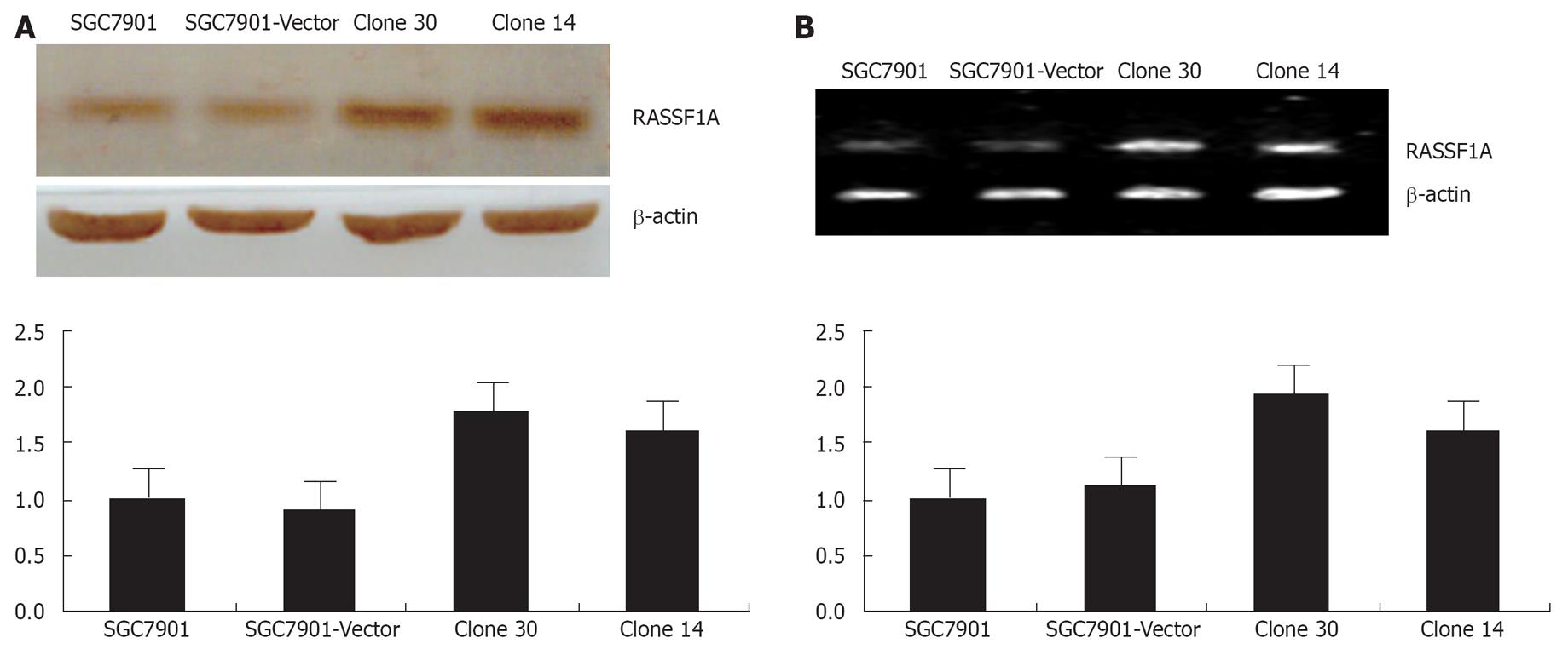
Figure 1 The over-expression of RASSF1A in gastric cancer cell line SGC7901.
Cells were stably transfected using Lipofectamine 2000 with RASSF1A or empty vector and were grown in RPMI containing G418 at 0.8 g/L. Resistant clones were selected separately and were measured by Western blotting and RT-PCR, β-actin was used as an internal loading control, and clone30 was used for further study. A: RASSF1A expression was analyzed by Western blotting; B: RASSF1A expression was analyzed by RT-PCR.
Figure 2 RASSF1A blocked gastric cancer cell line SGC7901 growth in vivo and in vitro.
A: Cell growth curve of gastric cancer cell line SGC7901 measured by MTT assay; B: RASSF1A induced gastric cancer cell line SGC7901 G1 arrest; C: RASSF1A inhibited the colony formation of SGC7901 cells measured by planting efficiency; D: RASSF1A inhibited the tumorigenicity of SGC7901 cells.
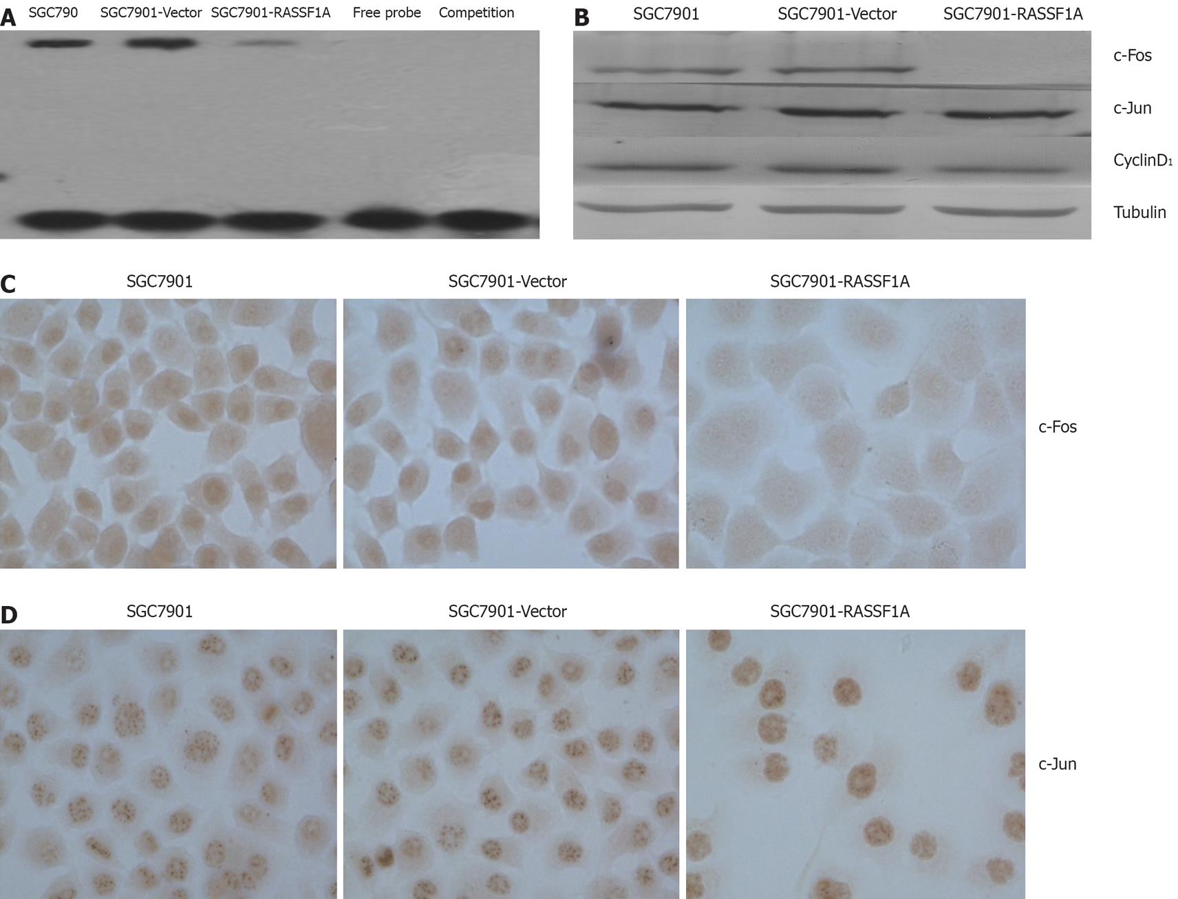
Figure 3 The over-expression of RASSF1A inhibited the AP-1 activity in gastric cancer cell line SGC7901.
A: The over-expression of RASSF1A inhibited the AP-1 DNA binding activity measured by EMSA; B: The over-expression of RASSF1A inhibited the c-Fos but not c-Jun expression in nuclear extract of SGC7901 cells. (Western blotting, DAB staining); C: The over-expression of RASSF1A inhibited the c-Fos expression in nuclear of SGC7901 cells (immunocytochemistry, DAB staining, × 400); D: The over-expression of RASSF1A had no influence on the c-Jun expression in nuclear of SGC7901 cells (immunocytochemistry, DAB staining, × 400).
- Citation: Deng ZH, Wen JF, Li JH, Xiao DS, Zhou JH. Activator protein-1 involved in growth inhibition by RASSF1A gene in the human gastric carcinoma cell line SGC7901. World J Gastroenterol 2008; 14(9): 1437-1443
- URL: https://www.wjgnet.com/1007-9327/full/v14/i9/1437.htm
- DOI: https://dx.doi.org/10.3748/wjg.14.1437


