Copyright
©2008 The WJG Press and Baishideng.
World J Gastroenterol. Mar 7, 2008; 14(9): 1346-1352
Published online Mar 7, 2008. doi: 10.3748/wjg.14.1346
Published online Mar 7, 2008. doi: 10.3748/wjg.14.1346
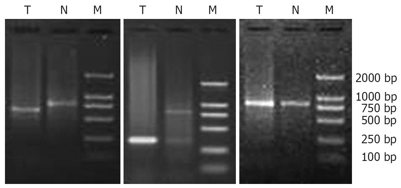
Figure 1 PCR amplified products of HBx-DNA.
The fragment sizes of PCR products range from 150 bp to 800 bp. M: Marker; T: Tumor; N: Non-tumor.
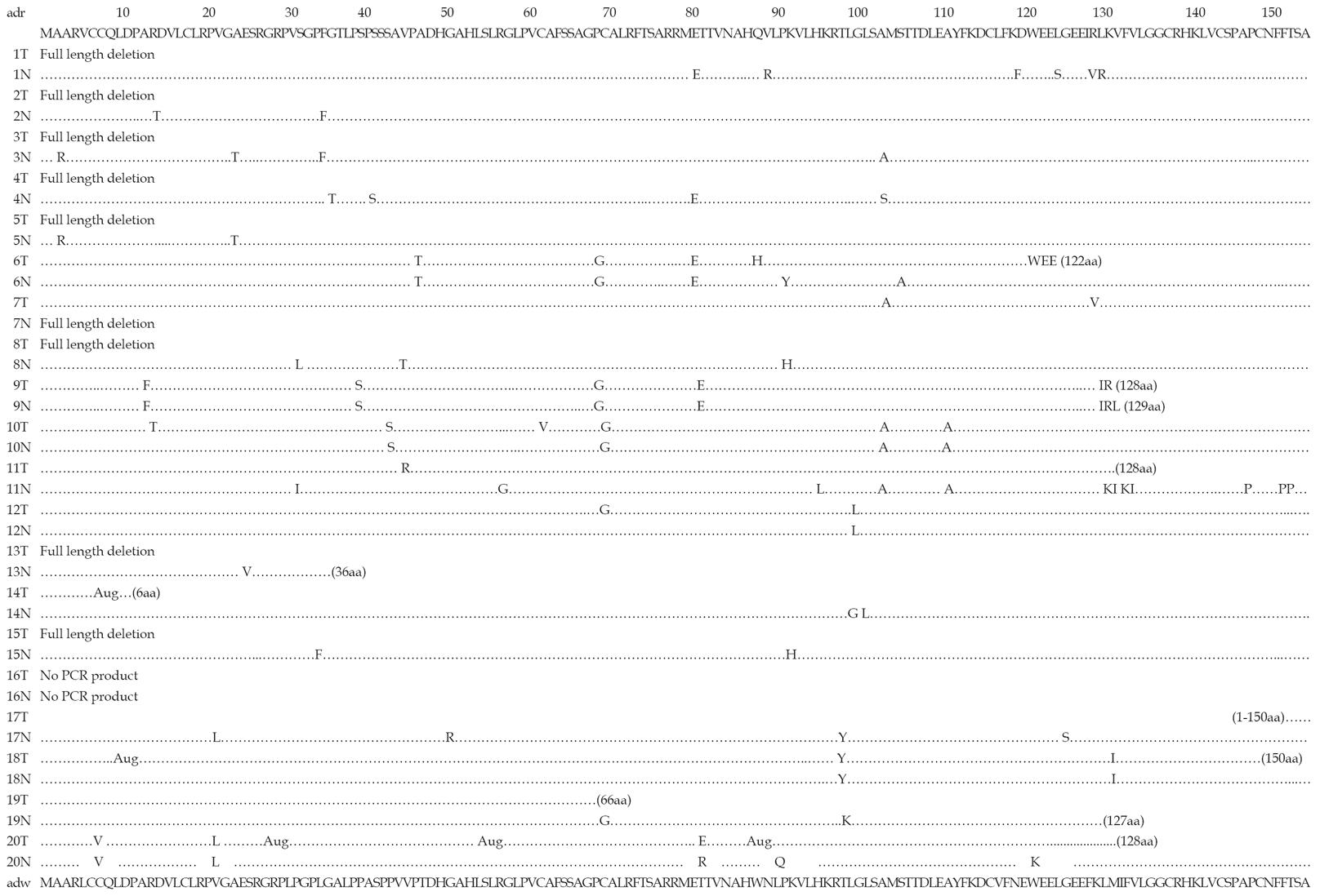
Figure 2 HBx sequencing in cancer and non-cancerous tissues from 20 HBV-associated HCC patients.
The amino acid sequences of HBV adr and adw subtypes are shown at the top or the bottom. Identical amino acid residues are represented by dots. The underlined amino acids were deduced from cellular flanking sequences. The frequencies of HBx point mutations were significantly lower in HCC than in the corresponding non-cancerous liver tissues (11/19 vs 18/19, P = 0.019). T: Tumor; N: Non-tumor. “…”: Represent the corresponding PCR amplificated base sequences.
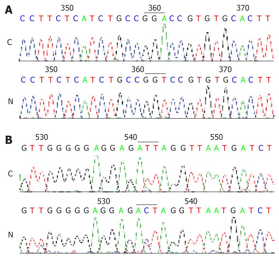
Figure 3 (A) GGA→GGT sense mutations at 67aa and (B) ATT→ACT mis-sense mutations at 127aa in non-cancerous liver tissues.
C: Control; N: Non-tumor.
Figure 4 Sequencing of HBx deletion mutation in HCC.
Figure 5 Sequencing of a specific integrated HBV at GA-rich region of 17p13 locus from HCC tissues of no.
2 patient. A: Tumor; B: Non-tumor.
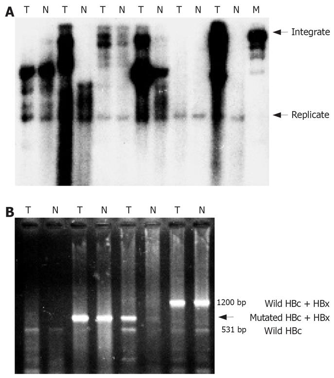
Figure 6 A: Detection of integrated HBx fragments in HCC tissues by Southern blot analysis; B: HBc and HBx fragment amplified by PCR.
M: Marker; T: Tumor; N: Non-tumor.
- Citation: Liu XH, Lin J, Zhang SH, Zhang SM, Feitelson MA, Gao HJ, Zhu MH. COOH-terminal deletion of HBx gene is a frequent event in HBV-associated hepatocellular carcinoma. World J Gastroenterol 2008; 14(9): 1346-1352
- URL: https://www.wjgnet.com/1007-9327/full/v14/i9/1346.htm
- DOI: https://dx.doi.org/10.3748/wjg.14.1346


