Copyright
©2008 The WJG Press and Baishideng.
World J Gastroenterol. Feb 28, 2008; 14(8): 1302-1304
Published online Feb 28, 2008. doi: 10.3748/wjg.14.1302
Published online Feb 28, 2008. doi: 10.3748/wjg.14.1302
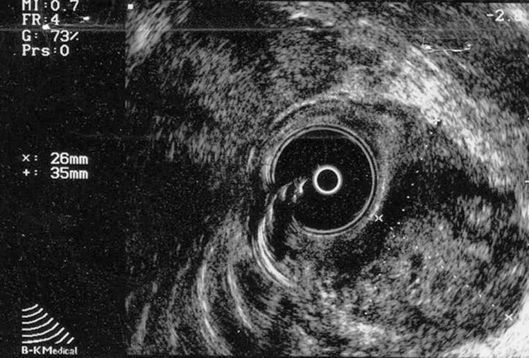
Figure 1 Transrectal ultrasound confirming a predominantly exophytic, heterogenous, hypoechoic submucosal mass (measuring 35 mm × 26 mm) on the lateral left rectal wall.
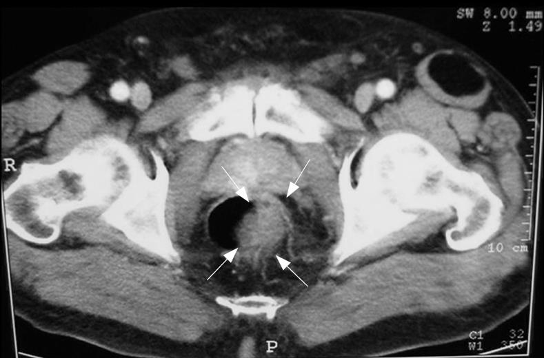
Figure 2 TC confirming the sonographic findings of the presence of a mass with a marked, irregular, eccentric thickening of the lateral left wall of the lower third of the rectum, but providing no evidence for either pelvic lymphadenopathy or distant metastasis.
- Citation: Grassi N, Cipolla C, Torcivia A, Mandalà S, Graceffa G, Bottino A, Latteri F. Gastrointestinal stromal tumour of the rectum: Report of a case and review of literature. World J Gastroenterol 2008; 14(8): 1302-1304
- URL: https://www.wjgnet.com/1007-9327/full/v14/i8/1302.htm
- DOI: https://dx.doi.org/10.3748/wjg.14.1302


