Copyright
©2008 The WJG Press and Baishideng.
World J Gastroenterol. Feb 28, 2008; 14(8): 1274-1279
Published online Feb 28, 2008. doi: 10.3748/wjg.14.1274
Published online Feb 28, 2008. doi: 10.3748/wjg.14.1274
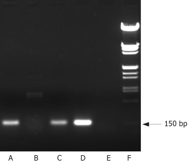
Figure 1 CNHK300 hTERT mRNA expression in various cell lines.
Telomerase hTERT mRNA expression was positive in cancer cell lines HepGII (A) and Hep3B (C), but not in the normal fibroblast cell line BJ (B) CNHK300 hTERT mRNA expression was detected in E1 transformed human embryonal kidney cell line HEK293 (D) as a positve control. E: Negative control, F: DNA marker.
Figure 2 Detection of E1A expression by Western blotting.
CNHK300 could express E1A in HepGII (A) and Hep3B (C) cells but not in the BJ human normal fibroblast cells (E), whereas wtAd5 did not show any selectivity in E1A expression, wtAd5 E1A expression was positive in Hep3B cells (D). E1A expression was detected in E1 transformed human embryonic kidney cell line HEK293 (B) as a positive control.
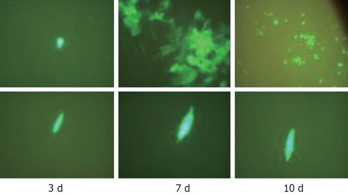
Figure 3 Proliferation selectivity of CNHK300-GFP in Hep3B (upper) and BJ (lower) under fluorescence microscope.
CNHK300-GFP could infect both BJ cells and Hep3B cells. But after days of infection, CNHK300-GFP virus could proliferate effectively in cancer cells and induced cytopathologic effect (CPE) at d 7, whereas in normal cell lines, CNHK300-GFP proliferation was attenuated, and BJ cells did not show CPE after days of infection.
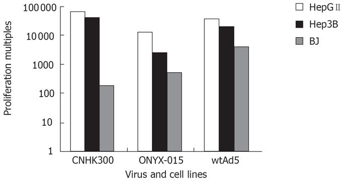
Figure 4 CNHK300, ONYX-015 and wtAd5 proliferation multiples in hepatocellular cancer cells HepGII, Hep3B and normal fibroblast cells BJ after 48 h virus infection.
CNHK300 replicated by 40 625 and 65 326 folds on tumor cell HepGII and Hep3B, similar to those of wtAd5. The CNHK300 replication (180-folds) was attenuated significantly as compared with wtAd5 (4000-folds) in BJ normal cell lines.
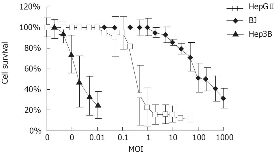
Figure 5 Hepatocellular cancer cells HepGII, Hep3B and normal fibroblast cells BJ survival curves after the cells were infected with CNHK300 at increasing MOIs.
CNHK300 caused significant cytolysis in HepGII and Hep3B tumor cell lines with a MOI of 0.5 pfu/cell and 0.0002 pfu/cell. For normal fibroblast cell lines BJ in contrast, cells infected with CNHK300 showed over 50% cell viability rate at the same time points with MOI of 100 pfu/cell.
- Citation: Li YM, Song ST, Jiang ZF, Zhang Q, Su CQ, Liao GQ, Qu YM, Xie GQ, Li MY, Ge FJ, Qian QJ. Telomerase-specific oncolytic virotherapy for human hepatocellular carcinoma. World J Gastroenterol 2008; 14(8): 1274-1279
- URL: https://www.wjgnet.com/1007-9327/full/v14/i8/1274.htm
- DOI: https://dx.doi.org/10.3748/wjg.14.1274


