Copyright
©2008 The WJG Press and Baishideng.
World J Gastroenterol. Feb 21, 2008; 14(7): 1108-1111
Published online Feb 21, 2008. doi: 10.3748/wjg.14.1108
Published online Feb 21, 2008. doi: 10.3748/wjg.14.1108
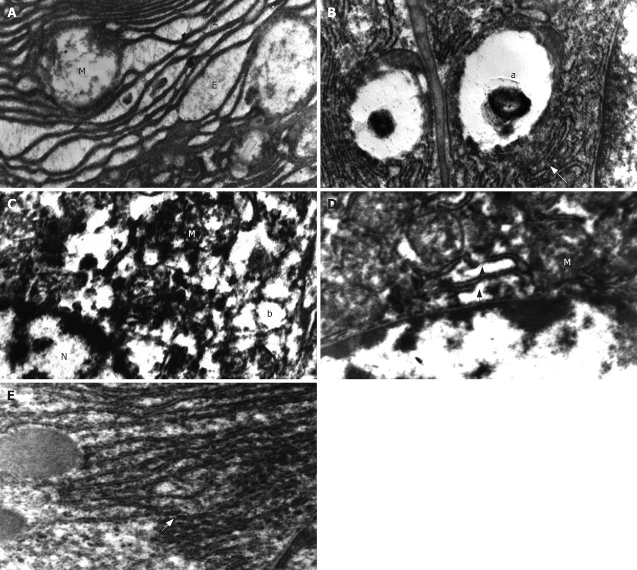
Figure 1 A: Electron micrograph showing irregular lamelar organizations and dilation of the RER (E) and swollen mitochondria (M) in the CCl4 group (× 15 000); B: Electron micrograph showing myelin figures in the SER (a) and breaks in the RER (arrow) in groupI (× 20 000); C: Electron micrograph showing the margination and clumping of the chromatin in the nucleus (N), mild swelling in a mitochondrion (M), mild dilation in the SER (b) and breaks in RER in group II (× 15 000); D: Electron micrograph showing mild swelling in the mitochondria (M) and breaks in the RER (arrow) in group II (× 20 000); E: Electron micrograph showing normal RER (arrow) in group II (× 20 000).
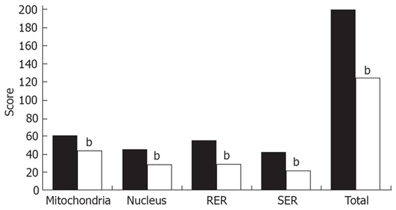
Figure 2 Ultrastructural injury scores in hepatocytes.
Individual and total organelle injury scores decreased significantly in UDCA-treated compared with saline-treated animals. bP < 0.001.
- Citation: Mas N, Tasci I, Comert B, Ocal R, Mas MR. Ursodeoxycholic acid treatment improves hepatocyte ultrastructure in rat liver fibrosis. World J Gastroenterol 2008; 14(7): 1108-1111
- URL: https://www.wjgnet.com/1007-9327/full/v14/i7/1108.htm
- DOI: https://dx.doi.org/10.3748/wjg.14.1108


