Copyright
©2008 The WJG Press and Baishideng.
World J Gastroenterol. Feb 7, 2008; 14(5): 805-807
Published online Feb 7, 2008. doi: 10.3748/wjg.14.805
Published online Feb 7, 2008. doi: 10.3748/wjg.14.805
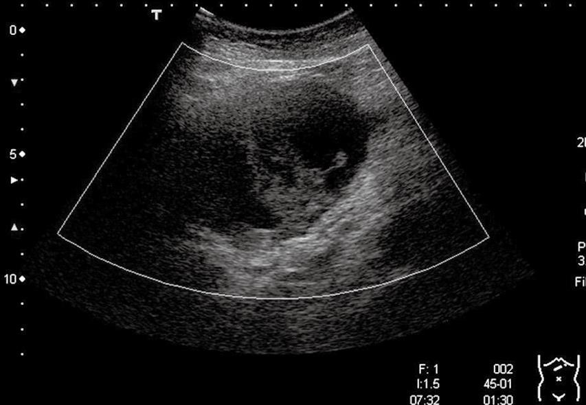
Figure 1 US image showing a heterogeneous echogenic cyst in S2, measuring 11.
0 cm × 8.0 cm and showing intracystic septations and structures.
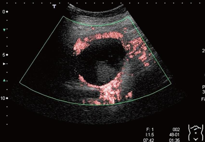
Figure 2 CEU images demonstrating absence of enhancement of the intracystic septations and structures and smooth of the enhanced cyst wall.
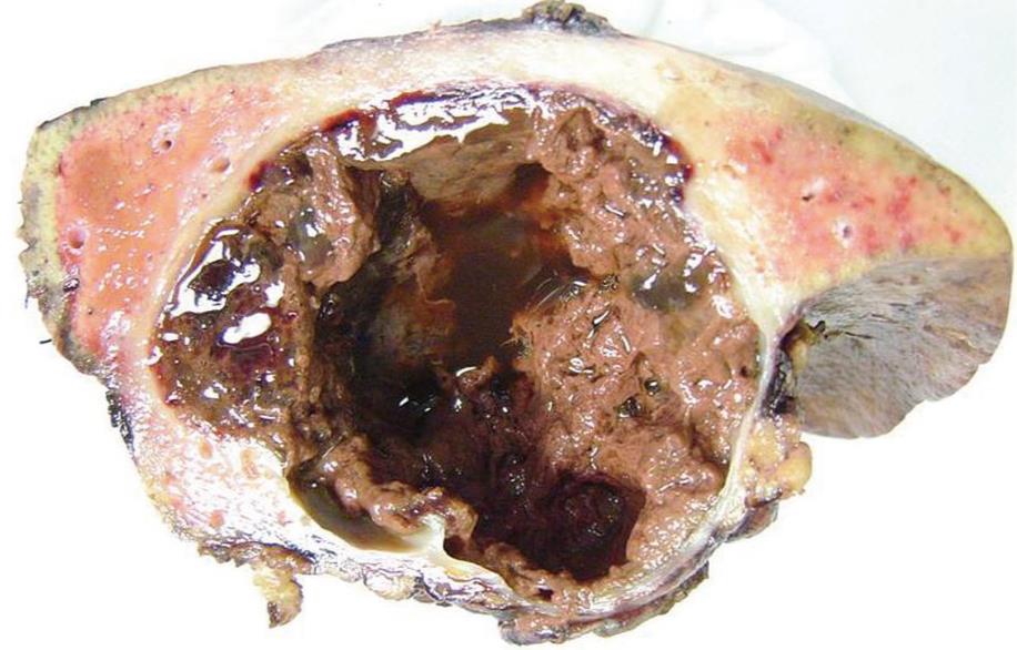
Figure 3 Histological examination showed that the intracystic structures were clots caused by the intracystic hemorrhage.
- Citation: Akiyama T, Inamori M, Saito S, Takahashi H, Yoneda M, Fujita K, Fujisawa T, Abe Y, Kirikoshi H, Kubota K, Ueda M, Tanaka K, Togo S, Ueno N, Shimada H, Nakajima A. Levovist ultrasonography imaging in intracystic hemorrhage of simple liver cyst. World J Gastroenterol 2008; 14(5): 805-807
- URL: https://www.wjgnet.com/1007-9327/full/v14/i5/805.htm
- DOI: https://dx.doi.org/10.3748/wjg.14.805


