Copyright
©2008 The WJG Press and Baishideng.
World J Gastroenterol. Feb 7, 2008; 14(5): 720-724
Published online Feb 7, 2008. doi: 10.3748/wjg.14.720
Published online Feb 7, 2008. doi: 10.3748/wjg.14.720
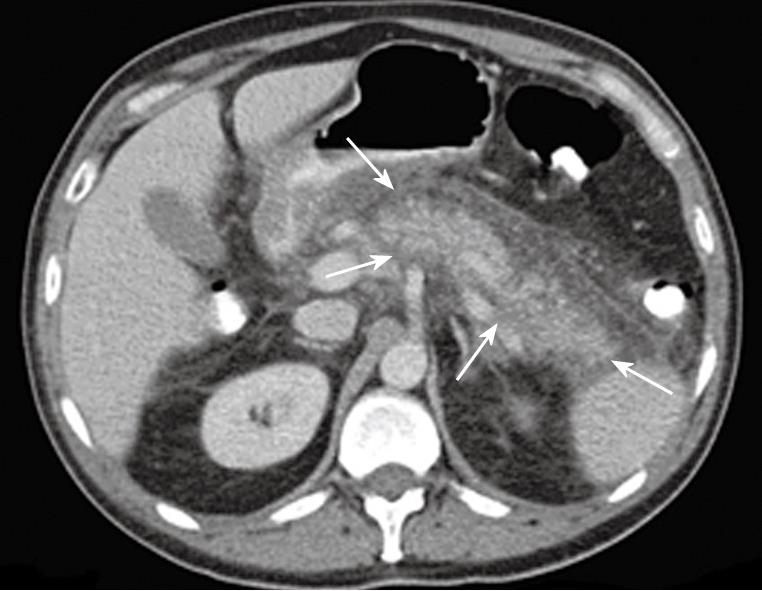
Figure 1 A CT image of a 23-year old female, who developed post-DBE pancreatitis (case 5), showing an inflamed body-tail region, with edema and fatty infiltration (arrows).
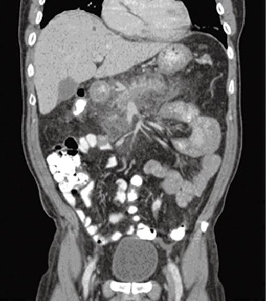
Figure 2 A transversal CT-image of a 54-year old male, who developed post-DBE pancreatitis (case 4), showing edema and fatty infiltration around the pancreas.
- Citation: Jarbandhan SV, Weyenberg SJV, Veer WMVD, Heine DG, Mulder CJ, Jacobs MA. Double balloon endoscopy associated pancreatitis: A description of six cases. World J Gastroenterol 2008; 14(5): 720-724
- URL: https://www.wjgnet.com/1007-9327/full/v14/i5/720.htm
- DOI: https://dx.doi.org/10.3748/wjg.14.720


