Copyright
©2008 The WJG Press and Baishideng.
World J Gastroenterol. Dec 21, 2008; 14(47): 7170-7174
Published online Dec 21, 2008. doi: 10.3748/wjg.14.7170
Published online Dec 21, 2008. doi: 10.3748/wjg.14.7170
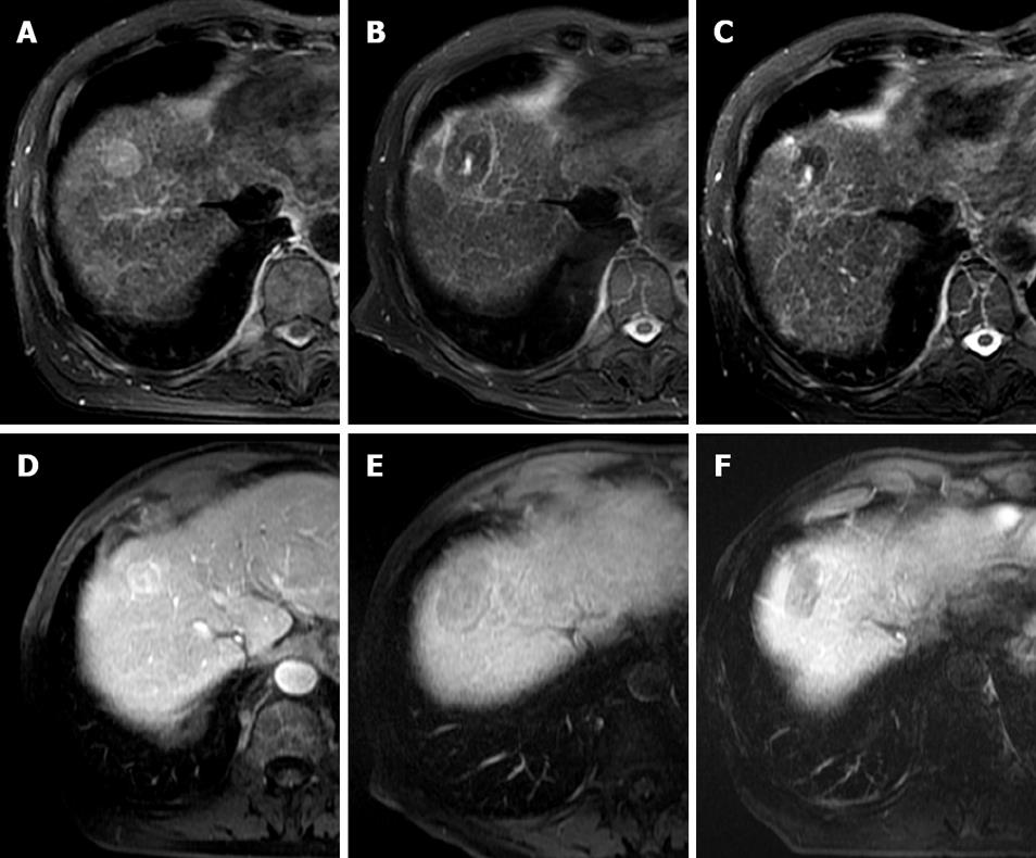
Figure 1 Typical MRI appearances pre and post laser ablation of a small HCC in a 73-year-old male patient with hepatitis C cirrhosis.
T2-weighted axial images demonstrate a high signal 2.5 cm segment VIII HCC (A). At one month post treatment (B) a typical low signal laser burn is demonstrated with high signal centre which has shrunk in size at 10 mo follow up (C). T1-weighted post contrast images of the same lesion demonstrate heterogeneous arterial enhancement pre-treatment (D), with non-enhancing scar at 1 mo (E) and persistent non-enhancement at 10 mo (F).
- Citation: Gough-Palmer AL, Gedroyc WMW. Laser ablation of hepatocellular carcinoma-A review. World J Gastroenterol 2008; 14(47): 7170-7174
- URL: https://www.wjgnet.com/1007-9327/full/v14/i47/7170.htm
- DOI: https://dx.doi.org/10.3748/wjg.14.7170


