Copyright
©2008 The WJG Press and Baishideng.
World J Gastroenterol. Dec 7, 2008; 14(45): 6924-6928
Published online Dec 7, 2008. doi: 10.3748/wjg.14.6924
Published online Dec 7, 2008. doi: 10.3748/wjg.14.6924
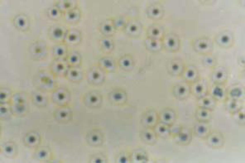
Figure 1 Culture of normal human gallbladder epithelial cells (HGBEC) (× 400).
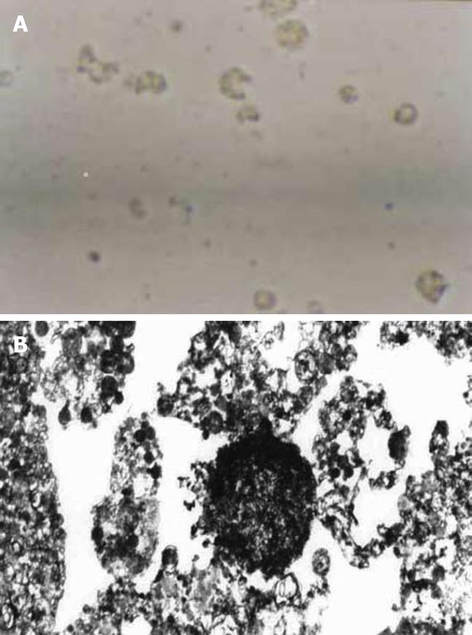
Figure 2 Four d after addition of enriched H pylori.
A: dead HGBEC (× 400); B: HGBEC characterized by karyopyknosis, vacuoles and incomplete cell structure (× 6000).
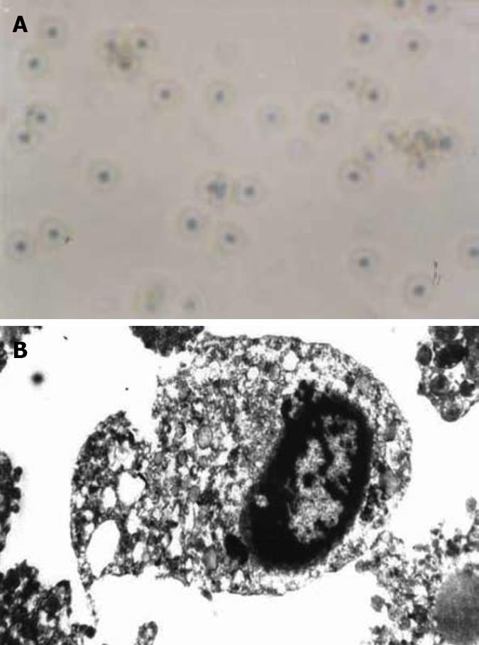
Figure 3 Four days after addition of H pylori sonicated extracts.
A: Most HGBEC were normal, however, a few HGBEC died (× 400); B: Basically normal HGBEC and some cellular plasm under the vacuole (× 6000).
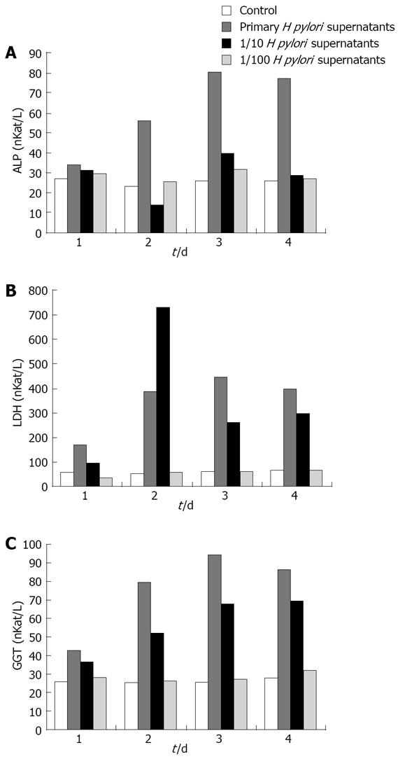
Figure 4 Effect of different concentrations of H pylori supernatants on ALP, LDH and GGT in the supernatants of HGBEC culture.
A: ALP; B: LDH; C: GGT.
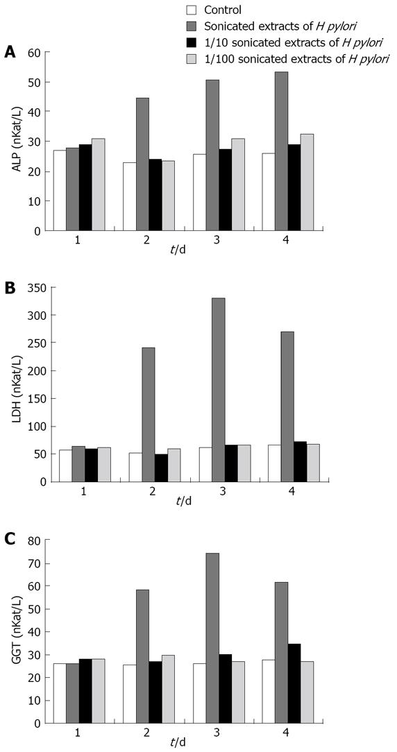
Figure 5 Effect of different concentrations of sonicated extracts of H pylori on HGBEC supernatant ALP, LDH and GGT.
A: ALP; B: LDH; C: GGT.
-
Citation: Chen DF, Hu L, Yi P, Liu WW, Fang DC, Cao H.
Helicobacter pylori damages human gallbladder epithelial cellsin vitro . World J Gastroenterol 2008; 14(45): 6924-6928 - URL: https://www.wjgnet.com/1007-9327/full/v14/i45/6924.htm
- DOI: https://dx.doi.org/10.3748/wjg.14.6924


