Copyright
©2008 The WJG Press and Baishideng.
World J Gastroenterol. Nov 28, 2008; 14(44): 6873-6875
Published online Nov 28, 2008. doi: 10.3748/wjg.14.6873
Published online Nov 28, 2008. doi: 10.3748/wjg.14.6873
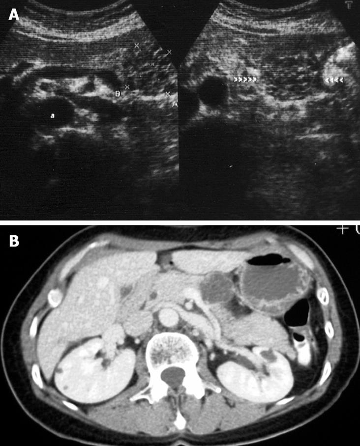
Figure 1 A well-circumscribed 35 mm polycystic lesion in the body of the pancreas, with thin septa within the lesion.
A: US scan demonstrating the polycystic tumour of the body of the pancreas; B: CT scan showing the cystic tumour with fine septa.
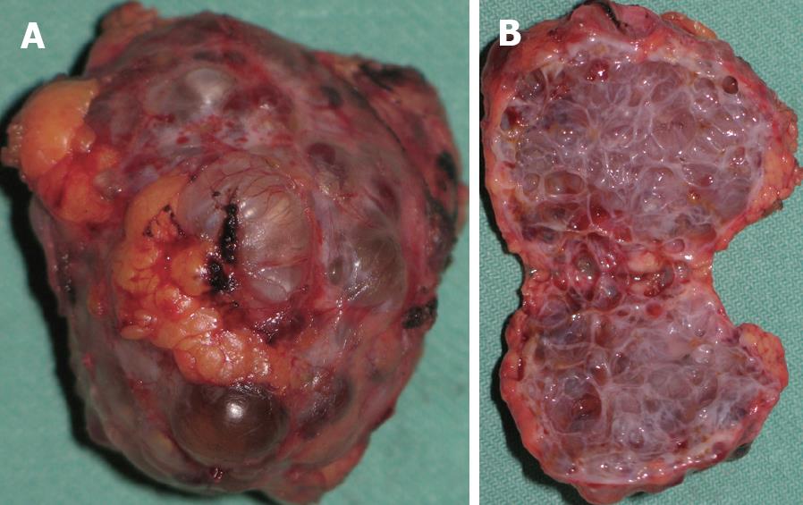
Figure 2 Tumour surrounded by normal pancreatic tissue.
A: The excised polycystic mass; B: The tumour after sectioning.
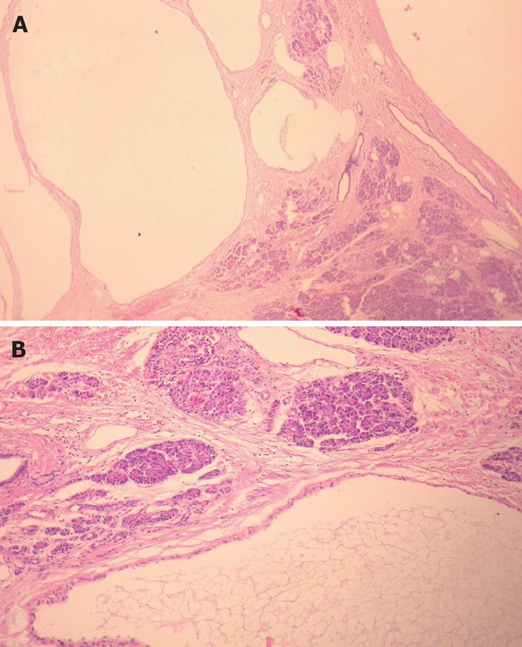
Figure 3 Microscopically all the sections (HE stain).
A: Vascular spaces of the pancreatic cystic Lymphangioma containing predominantly clear fluid with few erythrocytes or lymphocytes (× 13); B: The cysts and dilated lymphatics in the surrounding pancreatic tissue are lined with a thin endothelial layer (× 64).
- Citation: Colovic RB, Grubor NM, Micev MT, Atkinson HDE, Rankovic VI, Jagodic MM. Cystic lymphangioma of the pancreas. World J Gastroenterol 2008; 14(44): 6873-6875
- URL: https://www.wjgnet.com/1007-9327/full/v14/i44/6873.htm
- DOI: https://dx.doi.org/10.3748/wjg.14.6873


