Copyright
©2008 The WJG Press and Baishideng.
World J Gastroenterol. Nov 28, 2008; 14(44): 6867-6868
Published online Nov 28, 2008. doi: 10.3748/wjg.14.6867
Published online Nov 28, 2008. doi: 10.3748/wjg.14.6867
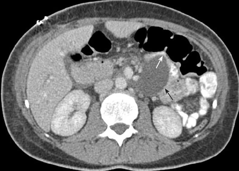
Figure 1 Contrast-enhanced CT showing the fluid collection (black arrow) compressing the efferent jejunal loop (white arrow) just distal to the gastrojejunostomy.
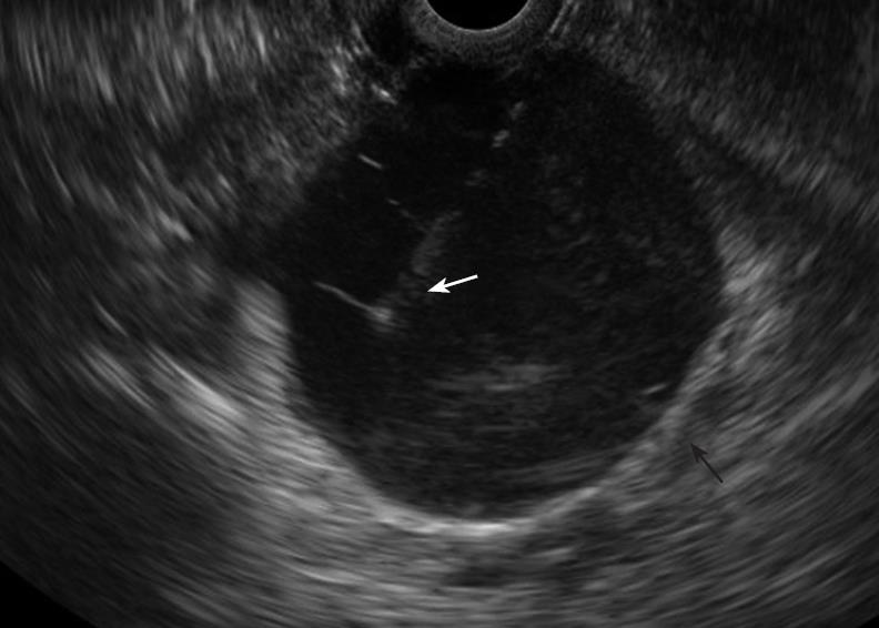
Figure 2 EUS showing the fluid collection (black arrow) with the needle in position for aspiration (white arrow).
- Citation: Jah A, Jamieson N, Huguet E, Griffiths W, Carroll N, Praseedom R. Endoscopic Ultrasound-guided drainage of an abdominal fluid collection following Whipple’s resection. World J Gastroenterol 2008; 14(44): 6867-6868
- URL: https://www.wjgnet.com/1007-9327/full/v14/i44/6867.htm
- DOI: https://dx.doi.org/10.3748/wjg.14.6867


