Copyright
©2008 The WJG Press and Baishideng.
World J Gastroenterol. Nov 7, 2008; 14(41): 6376-6381
Published online Nov 7, 2008. doi: 10.3748/wjg.14.6376
Published online Nov 7, 2008. doi: 10.3748/wjg.14.6376
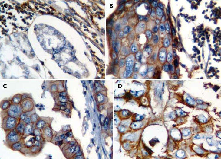
Figure 1 Representative example demonstrating the immunohistochemical staining of CD44 molecule in gastric adenocarcinoma tissue.
A: Negative CD44 staining in adeno-carcinoma glands and positive staining in the background lymphocytes (× 40); B: Membranous expression of CD44 in carcinoma cells with the stained lymphocytes as internal positive control in the background (× 40); C: Positive membranous staining of CD44 in carcinoma cells (× 40); D: Membranous and cytoplasmic expression of CD44 in carcinoma cells (× 40).
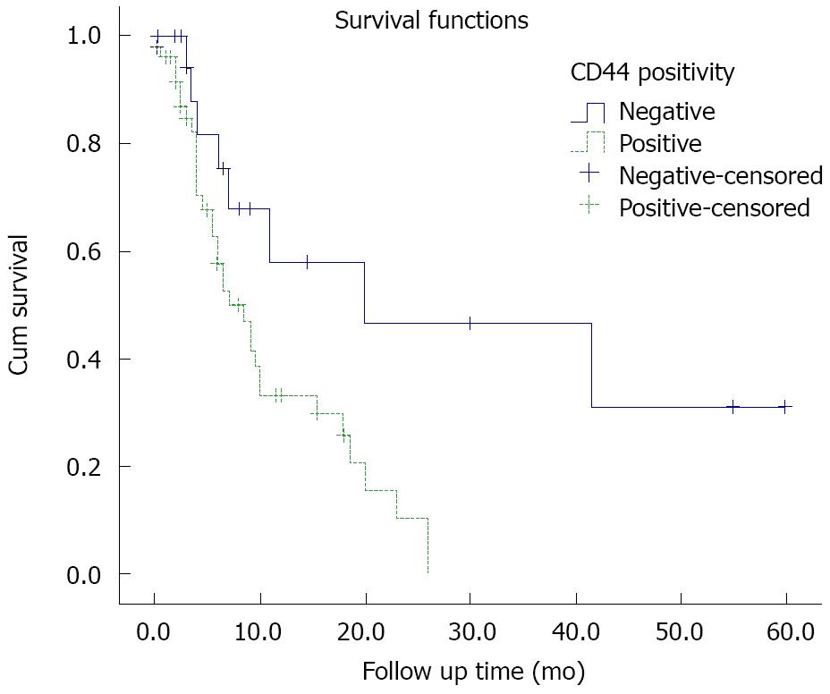
Figure 2 Kaplan-Meier curves of overall survival for CD44-positive and -negative gastric cancer cases.
- Citation: Ghaffarzadehgan K, Jafarzadeh M, Raziee HR, Sima HR, Esmaili-Shandiz E, Hosseinnezhad H, Taghizadeh-Kermani A, Moaven O, Bahrani M. Expression of cell adhesion molecule CD44 in gastric adenocarcinoma and its prognostic importance. World J Gastroenterol 2008; 14(41): 6376-6381
- URL: https://www.wjgnet.com/1007-9327/full/v14/i41/6376.htm
- DOI: https://dx.doi.org/10.3748/wjg.14.6376


