Copyright
©2008 The WJG Press and Baishideng.
World J Gastroenterol. Jan 28, 2008; 14(4): 622-626
Published online Jan 28, 2008. doi: 10.3748/wjg.14.622
Published online Jan 28, 2008. doi: 10.3748/wjg.14.622
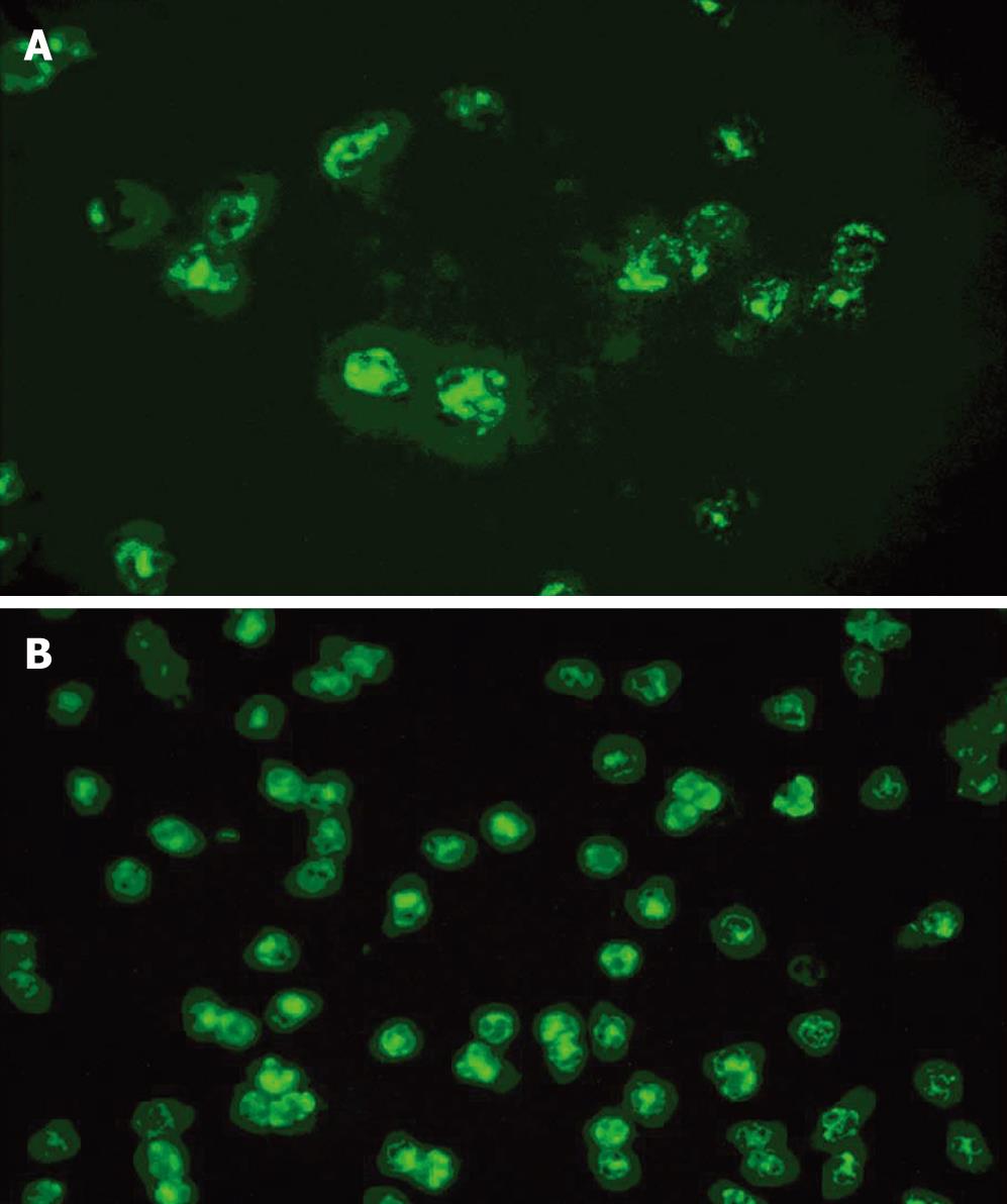
Figure 1 Strong cytoplasmic (A) and nuclear (B) staining patterns of ANCA (C-ANCA and P-ANCA) detected by indirect IIF.
Serum samples from patients were diluted and incubated with primary antibody, then with FITC-labeled affinity-purified goat anti-human IgG. The results were observed under fluorescence microscope.
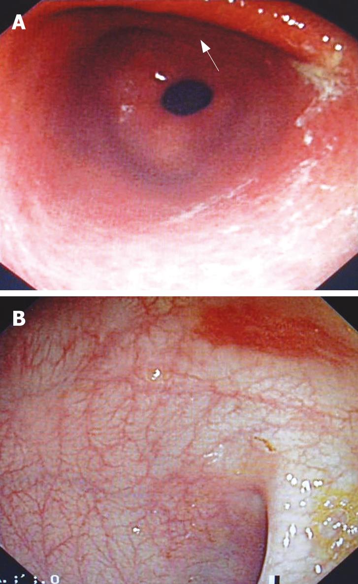
Figure 2 Gastroendoscopy showing edema and congestion in the pyloric region (A) and several petechiae scattering throughout the colon (B).
- Citation: Zhang Y, Wu YK, Ciorba MA, Ouyang Q. Significance of antineutrophil cytoplasmic antibody in adult patients with Henoch-Schönlein purpura presenting mainly with gastrointestinal symptoms. World J Gastroenterol 2008; 14(4): 622-626
- URL: https://www.wjgnet.com/1007-9327/full/v14/i4/622.htm
- DOI: https://dx.doi.org/10.3748/wjg.14.622


