Copyright
©2008 The WJG Press and Baishideng.
World J Gastroenterol. Jan 28, 2008; 14(4): 493-497
Published online Jan 28, 2008. doi: 10.3748/wjg.14.493
Published online Jan 28, 2008. doi: 10.3748/wjg.14.493
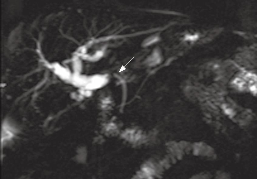
Figure 1 Magnetic resonance cholagiopancreatography (MRCP) image of the biliary tract of a patient that developed abnormal liver enzymes 8 mo after liver transplantation.
The MRCP showed a severe biliary stricture at the anastomotic site (arrow).
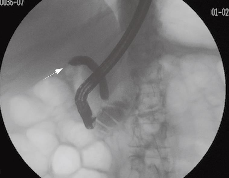
Figure 2 Endoscopic retrograde cholangiography (ERC) in a patient that developed pruritus and cholestasis 6 mo after liver transplantation.
The ERC showed a severe anastomosis stricture (arrow). This stricture was successfully managed with biliary balloon dilation and stent placement.
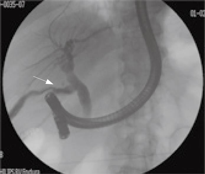
Figure 3 Endoscopic retrograde cholangiography (ERC) image of a biliary leak in a patient after a liver transplantation.
The arrow is pointing to the area of extravasation of contrast from the biliary tree.
- Citation: Londoño MC, Balderramo D, Cárdenas A. Management of biliary complications after orthotopic liver transplantation: The role of endoscopy. World J Gastroenterol 2008; 14(4): 493-497
- URL: https://www.wjgnet.com/1007-9327/full/v14/i4/493.htm
- DOI: https://dx.doi.org/10.3748/wjg.14.493


