Copyright
©2008 The WJG Press and Baishideng.
World J Gastroenterol. Oct 21, 2008; 14(39): 6096-6099
Published online Oct 21, 2008. doi: 10.3748/wjg.14.6096
Published online Oct 21, 2008. doi: 10.3748/wjg.14.6096
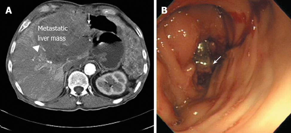
Figure 1 Diagnostic evaluations of recurrent gastrointestinal stromal tumors before starting sunitinib.
A: In computed tomography, arrowhead and arrow show metastatic liver mass and colon invasion, respectively. B: In the colonoscopic finding, arrow shows a protruding mass invading from the external lumen.
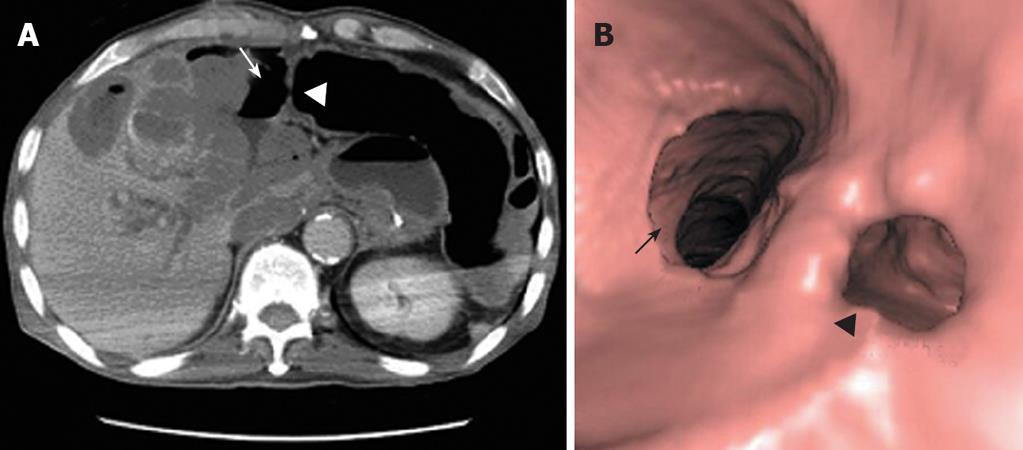
Figure 2 Diagnostic evaluation of perforating colon in present symptomatic patient during sunitinib treatment.
A: In computed tomography, arrow and arrowhead show intraperitoneal free air and the site of colon perforation, respectively. B: In 3-dimensional reconstruction, arrow and arrowhead show the colon lumen and the site of perforation, respectively..
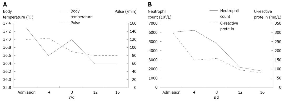
Figure 3 Changes in body temperature and pulse (A) and in neutrophil counts and C-reactive protein (B) of the patient.
- Citation: Hur H, Park AR, Jee SB, Jung SE, Kim W, Jeon HM. Perforation of the colon by invading recurrent gastrointestinal stromal tumors during sunitinib treatment. World J Gastroenterol 2008; 14(39): 6096-6099
- URL: https://www.wjgnet.com/1007-9327/full/v14/i39/6096.htm
- DOI: https://dx.doi.org/10.3748/wjg.14.6096


