Copyright
©2008 The WJG Press and Baishideng.
World J Gastroenterol. Oct 14, 2008; 14(38): 5927-5929
Published online Oct 14, 2008. doi: 10.3748/wjg.14.5927
Published online Oct 14, 2008. doi: 10.3748/wjg.14.5927
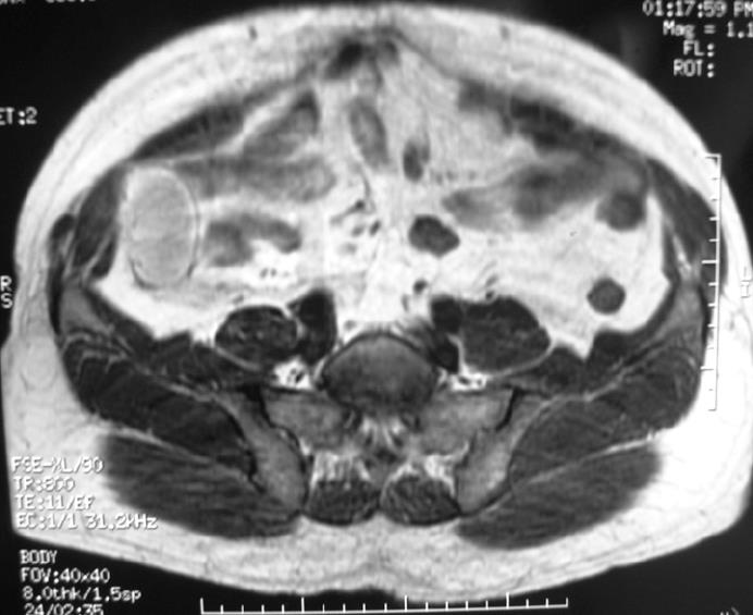
Figure 1 T1-weighted magnetic resonance image (MRI) demonstrating a high-intensity mass.
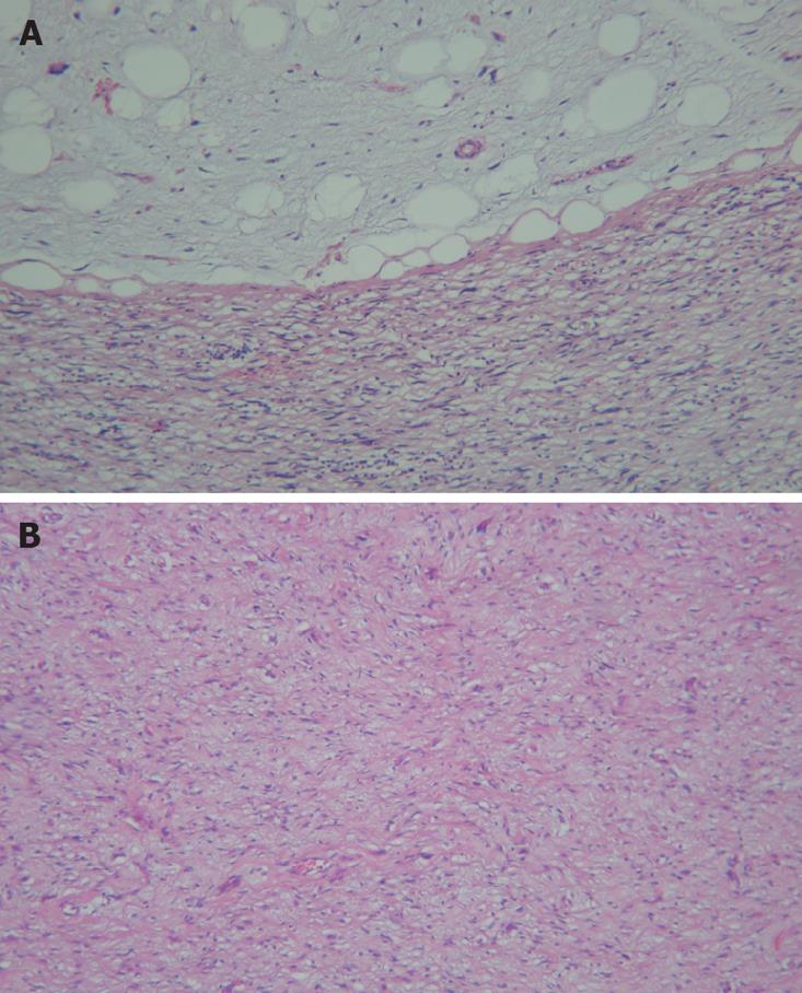
Figure 2 Well-differentiated tumor shows numerous lipoblasts and the dedifferentiated tumor resembles pleomorphic, spindle cells with hyperchromatic nuclei (HE, × 100) in the area revealing an abrupt transition from the well-differentiated to the dedifferentiated tumor (A), and dedifferentiated tumor shows low cellularity, fusiform cells with small hyperchromatic nuclei, and abundant collogen in the low grade fibrosarcoma area (B) (HE, × 100).
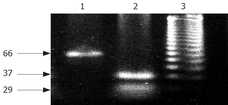
Figure 3 Amplification of the BstU1 within exon 4 produces a 66 bp long segment while cleavage results in 37 bp and 29 bp long fragments.
Lane 1: No mutation of p53 gene in well-differentiated tumor; lane 2: Mutation of p53 gene in dedifferentiated tumor; lane 3: DNA marker.
- Citation: Karaman A, Kabalar ME, Özcan &, Koca T, Binici DN. Intraperitoneal dedifferentiated liposarcoma: A case report. World J Gastroenterol 2008; 14(38): 5927-5929
- URL: https://www.wjgnet.com/1007-9327/full/v14/i38/5927.htm
- DOI: https://dx.doi.org/10.3748/wjg.14.5927


