Copyright
©2008 The WJG Press and Baishideng.
World J Gastroenterol. Oct 14, 2008; 14(38): 5920-5923
Published online Oct 14, 2008. doi: 10.3748/wjg.14.5920
Published online Oct 14, 2008. doi: 10.3748/wjg.14.5920
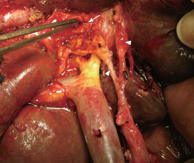
Figure 1 Skeletonization for regional lymphadenectomy with removal of neural and lymphoid tissues from hepatoduodenal ligament and liver hilus in a type II hilar cholangiocarcinoma.
A too-tight dissection around the right and left hepatic arteries (arrow heads) is shown. Left hepatic duct (arrow) is being prepared for bilio-enteric anastomosis.
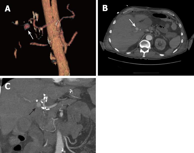
Figure 2 MDCTA.
A: Arterial phase image precisely depicts a large pseudoaneurysm originating from the right hepatic artery (arrow). B and C: Jejunum Y-limb did not show a demonstrable communication with the arterial sac in cross-sectional and coronal views, respectively.
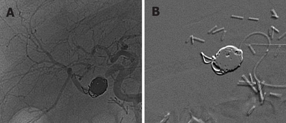
Figure 3 Patency of the hepatic artery and its branches, and the occlusion of the pseudoaneurysm.
A: Embolization of the pseudoaneurysm using microcoils to stop the inflow into the pseudoaneurysmal lumen with patency of the hepatic artery and its branches. B: Subtracted angiogram after right hepatic artery pseudoaneurysm occlusion with stainless steel coils.
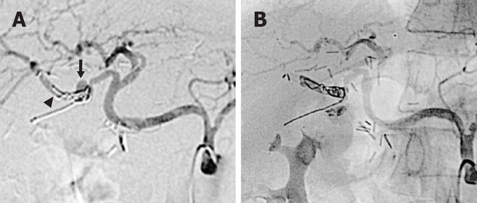
Figure 4 Partial revascularization of the pseudoaneurysm and TAE of the right hepatic artery.
A: Angiography 3 wk after the embolization of the right hepatic artery pseudoaneurysm with a partial revascularization of the arterial sac (arrow) and a distal migration of coils (arrow head). A partial enteric migration of microcoils after the first and selective embolization suggests an arterio-enteric fistula origin. B: TAE of the right hepatic artery with stainless steel coils and polyvinyl alcohol particles. No distal arterial flow can be shown.
- Citation: Briceño J, Naranjo &, Ciria R, Sánchez-Hidalgo JM, Zurera L, López-Cillero P. Late hepatic artery pseudoaneurysm: A rare complication after resection of hilar cholangiocarcinoma. World J Gastroenterol 2008; 14(38): 5920-5923
- URL: https://www.wjgnet.com/1007-9327/full/v14/i38/5920.htm
- DOI: https://dx.doi.org/10.3748/wjg.14.5920


