Copyright
©2008 The WJG Press and Baishideng.
World J Gastroenterol. Oct 7, 2008; 14(37): 5769-5772
Published online Oct 7, 2008. doi: 10.3748/wjg.14.5769
Published online Oct 7, 2008. doi: 10.3748/wjg.14.5769
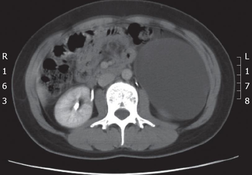
Figure 1 Contrast-enhanced CT of the abdomen showing a 12 cm × 6.
5 cm homogenous cystic mass in the retroperitoneal space with medial displacement of the descending colon.
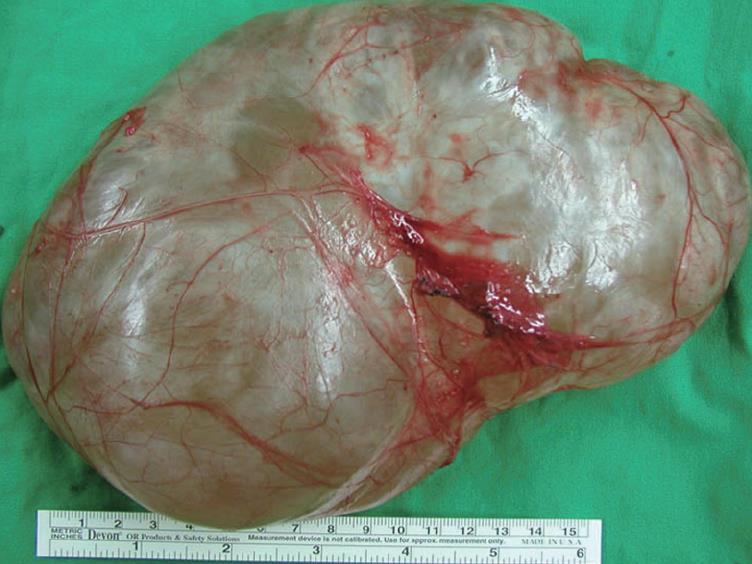
Figure 2 Photograph of the resected mass measuring 20 cm × 14 cm × 6 cm in size.
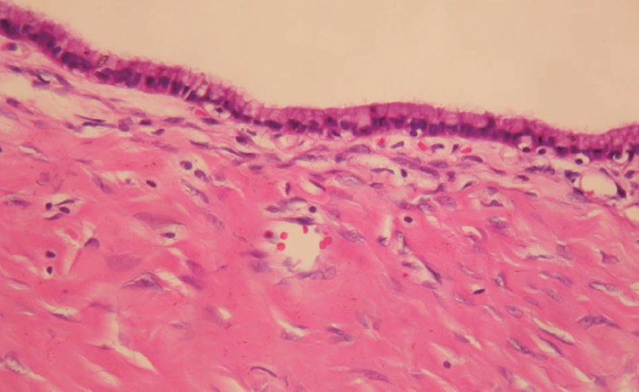
Figure 3 Photomicrograph showing a single layer of mucin-producing columnar epithelium with underlying fibrous connective tissue (HE, × 100).
- Citation: Yan SL, Lin H, Kuo CL, Wu HS, Huang MH, Lee YT. Primary retroperitoneal mucinous cystadenoma: Report of a case and review of the literature. World J Gastroenterol 2008; 14(37): 5769-5772
- URL: https://www.wjgnet.com/1007-9327/full/v14/i37/5769.htm
- DOI: https://dx.doi.org/10.3748/wjg.14.5769


