Copyright
©2008 The WJG Press and Baishideng.
World J Gastroenterol. Sep 28, 2008; 14(36): 5564-5569
Published online Sep 28, 2008. doi: 10.3748/wjg.14.5564
Published online Sep 28, 2008. doi: 10.3748/wjg.14.5564
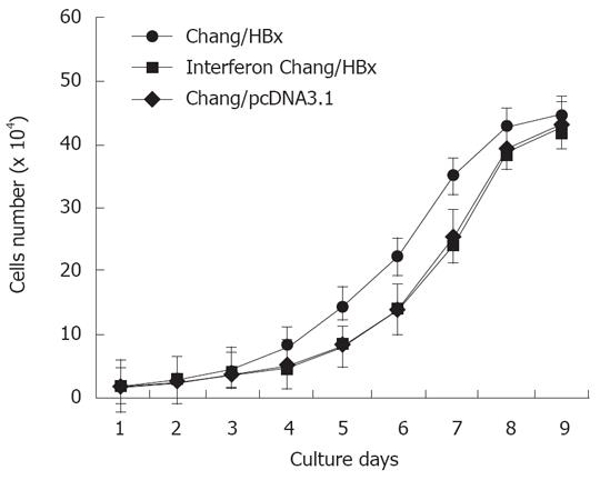
Figure 1 The growth curve of Chang/HBx cells shows a shift to the left from that of Chang/pcDNA3.
1 and IFN-α-Chang/HBx cells.
Figure 2 Cell proliferative ability comparisons.
The average colony-forming efficiency of Chang/HBx (A) was 29.3 ± 4.5, which was significantly different from that of Chang/pcDNA3.1 (B) (12.8 ± 2.6) and IFN-α-Chang/HBx (C) cells (13.5 ± 2.3) (P < 0.05).
Figure 3 HBx promotes the invasive capacity of Chang liver cells in vitro.
Wound-healing assays were performed at 0 and 24 h and observed under a phase-contrast microscope (× 100). A: Chang liver cells with pcDNA3.1-HBx were disrupted, and wound healing occurred after 24 h; B: IFN-α-Chang/HBx cells were disrupted, and the wound did not heal completely after 24 h; C: Chang cells with pcDNA3.1 were disrupted, and the wound did not heal completely after 24 h.
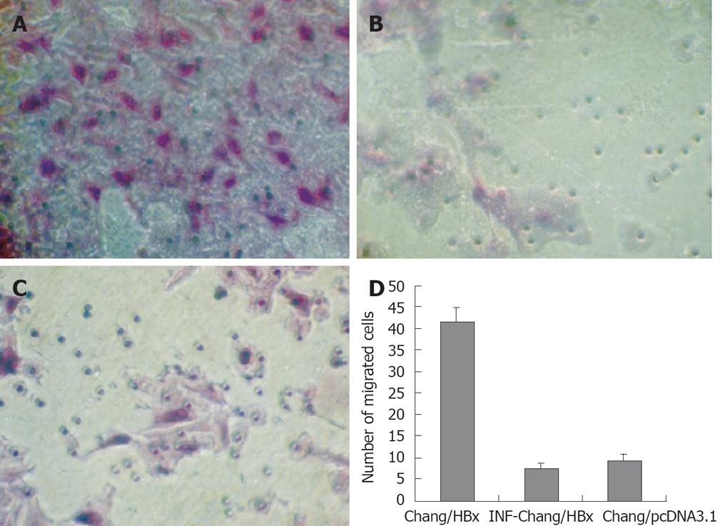
Figure 4 HBx promotes the invasive capacity of Chang liver cells in vitro.
A: Chang cells transfected with pcDNA3.1-HBx invaded through the Matrigel were counted on the underside of the transwell filter and compared with Chang/HBx cells (B) and control cells Chang/pcDNA3.1 (C) (× 200). There was no difference between IFN-α-Chang/HBx cells and Chang/pcDNA3.1; D: The average number of migrated cells per site seen under a high-power microscope (× 400) was 41.6 ± 3.1 for the transfected Chang/HBx cells and 7.4 ± 1.2 for IFN-α-Chang/HBx cells and 9.2 ± 1.6 for the control Chang/pcDNA3.1 cells.
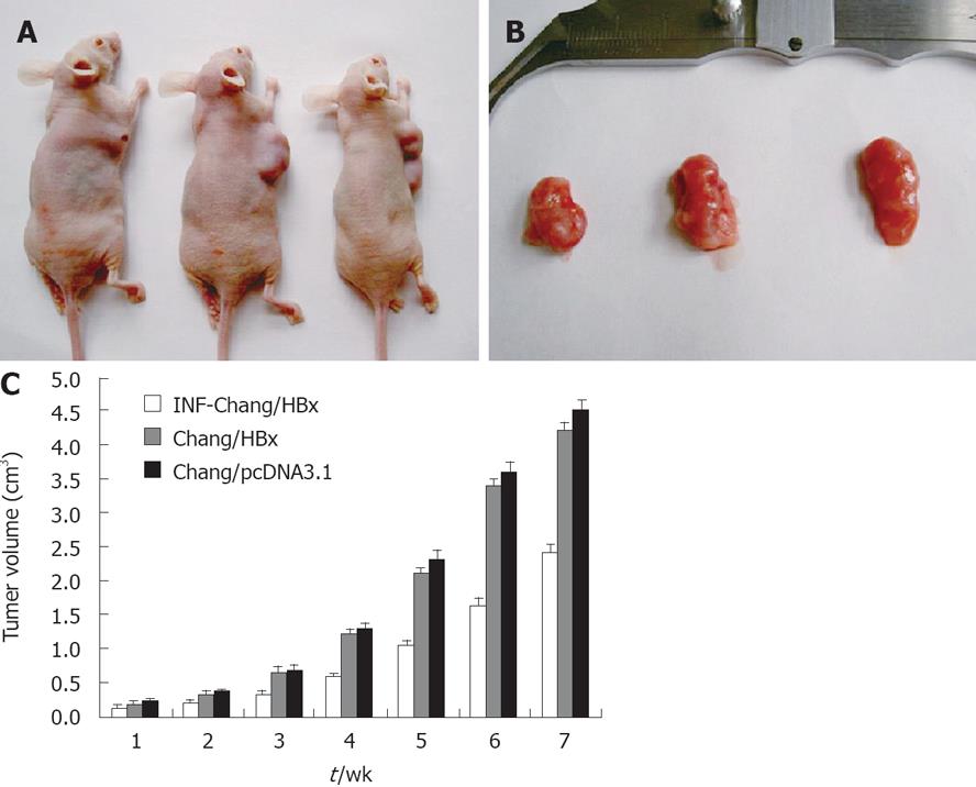
Figure 5 A: A nude mouse migration model with pcDNA3.
1-HBx Chang cells, IFN-α-Chang/HBx cells and Chang/pcDNA3.1 cells were constructed on the 7th wk; B: The volume of neoplasms in the three groups were observed. The sizes of the hepatomas from Chang/pcDNA3.1 and IFN-α-Chang/HBx injected nude mice were obviously larger than the tumors of Chang/HBx injected mice; C: Neoplasm growth in the Chang/HBx-inoculated nude mice compared between IFN-α-Chang/HBx and the control Chang/pcDNA3.1-inoculated mice.
- Citation: Yang JQ, Pan GD, Chu GP, Liu Z, Liu Q, Xiao Y, Yuan L. Interferon-alpha restrains growth and invasive potential of hepatocellular carcinoma induced by hepatitis B virus X protein. World J Gastroenterol 2008; 14(36): 5564-5569
- URL: https://www.wjgnet.com/1007-9327/full/v14/i36/5564.htm
- DOI: https://dx.doi.org/10.3748/wjg.14.5564


