Copyright
©2008 The WJG Press and Baishideng.
World J Gastroenterol. Sep 21, 2008; 14(35): 5481-5483
Published online Sep 21, 2008. doi: 10.3748/wjg.14.5481
Published online Sep 21, 2008. doi: 10.3748/wjg.14.5481
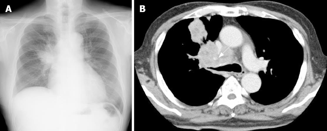
Figure 1 A: Chest X-ray revealed a large abnormal shadow in the right upper lobe; B: A computed tomography (CT) of the chest demonstrated a large lung tumor in the right upper lobe obstructing the right upper bronchus.
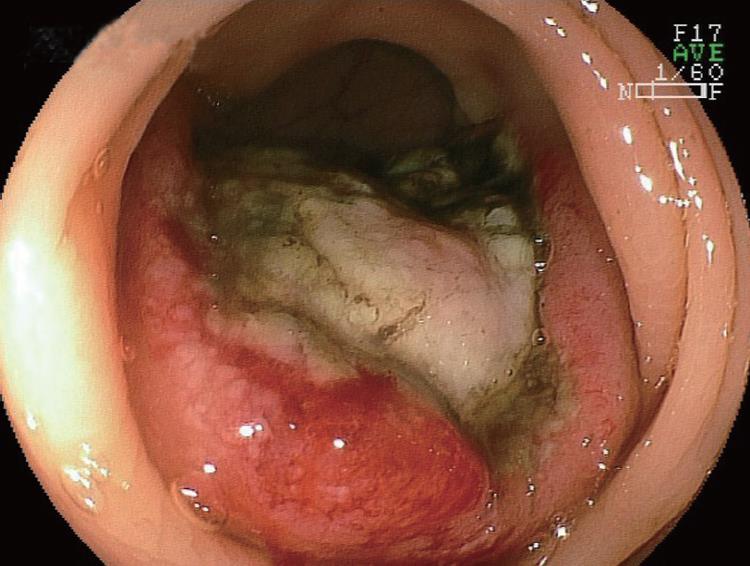
Figure 2 Endoscopic appearance of the descending colon.
A large protruding lesion with central ulceration, about 40 mm in diameter, was seen.
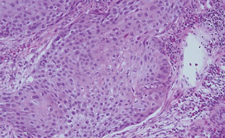
Figure 3 Histological examination of the biopsy specimen obtained from the colonic protruding lesion revealed tumor cells diagnosed as SCC (× 50).
- Citation: Hirasaki S, Suzuki S, Umemura S, Kamei H, Okuda M, Kudo K. Asymptomatic colonic metastases from primary squamous cell carcinoma of the lung with a positive fecal occult blood test. World J Gastroenterol 2008; 14(35): 5481-5483
- URL: https://www.wjgnet.com/1007-9327/full/v14/i35/5481.htm
- DOI: https://dx.doi.org/10.3748/wjg.14.5481


