Copyright
©2008 The WJG Press and Baishideng.
World J Gastroenterol. Sep 21, 2008; 14(35): 5478-5480
Published online Sep 21, 2008. doi: 10.3748/wjg.14.5478
Published online Sep 21, 2008. doi: 10.3748/wjg.14.5478
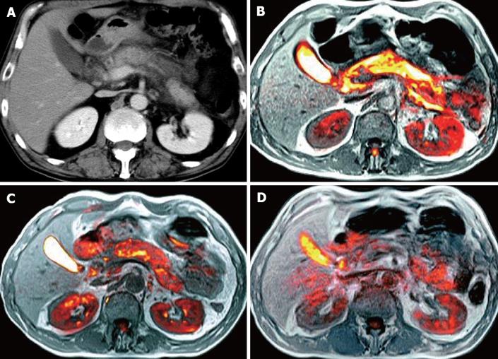
Figure 1 A CT scan at admission revealing enlarged pancreas complicated by acute multiple ascites (A), a fusion image at admission (B), on days 10 (C) and 50 (D) showing bright signals in the whole pancreas and ascites around it, diminished pancreatic enlargement and slightly decreased signal-intensity, as well as disappearance of all signs of acute pancreatitis, respectively.
- Citation: Shinya S, Sasaki T, Nakagawa Y, Guiquing Z, Yamamoto F, Yamashita Y. Acute pancreatitis successfully diagnosed by diffusion-weighted imaging: A case report. World J Gastroenterol 2008; 14(35): 5478-5480
- URL: https://www.wjgnet.com/1007-9327/full/v14/i35/5478.htm
- DOI: https://dx.doi.org/10.3748/wjg.14.5478


