Copyright
©2008 The WJG Press and Baishideng.
World J Gastroenterol. Sep 14, 2008; 14(34): 5349-5352
Published online Sep 14, 2008. doi: 10.3748/wjg.14.5349
Published online Sep 14, 2008. doi: 10.3748/wjg.14.5349
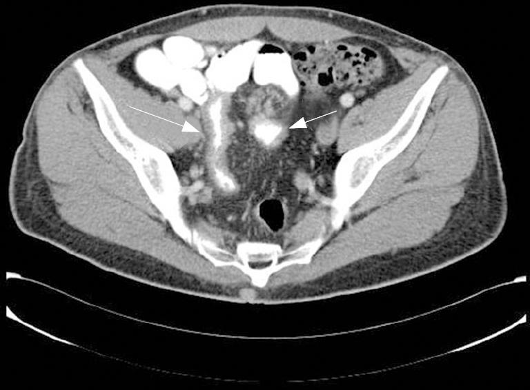
Figure 1 CT-scan showing thickening of the distal ileum (long arrow) with an adjacent solid mass (short arrow), suggesting a carcinoid tumour.
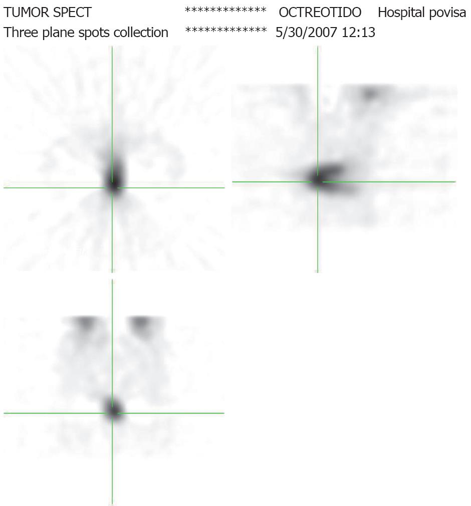
Figure 2 Octeotride-SPECT images showing pathological uptake in the ileal thickened loop at CT-scan.
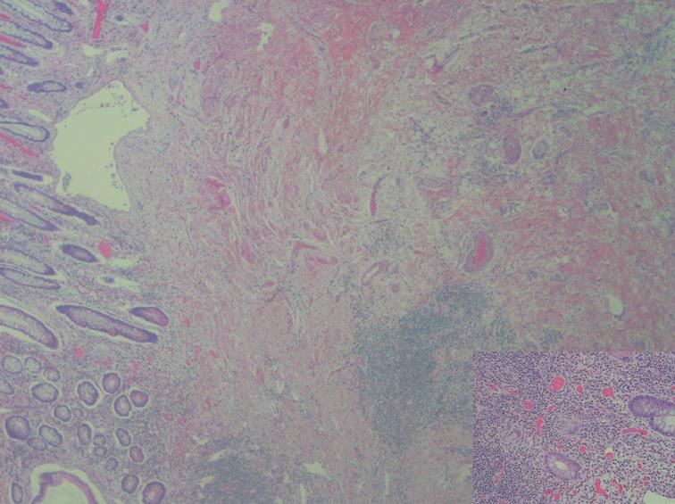
Figure 3 CD showing marked transmural inflammatory changes (involving the walls of veins and arteries): edema, lymphatic dilatation, hyperplasia of the muscularis mucosae, fibrosis (“obliterative muscularization”), and epithelioid granuloma.
- Citation: Fernandez A, Tabuenca O, Peteiro A. A “false positive” octreoscan in ileal Crohn’s disease. World J Gastroenterol 2008; 14(34): 5349-5352
- URL: https://www.wjgnet.com/1007-9327/full/v14/i34/5349.htm
- DOI: https://dx.doi.org/10.3748/wjg.14.5349


