Copyright
©2008 The WJG Press and Baishideng.
World J Gastroenterol. Sep 7, 2008; 14(33): 5162-5175
Published online Sep 7, 2008. doi: 10.3748/wjg.14.5162
Published online Sep 7, 2008. doi: 10.3748/wjg.14.5162
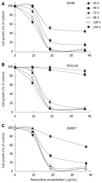
Figure 1 Time and dose dependent effect of roscovitine on the proliferation of human colorectal cancer cell lines.
Human colorectal cancer cell lines SW48 (A), SW1116 (B) and SW837 (C) were plated (27 × 103 cells/well) into 96-well plates and incubated at 37°C in a non-CO2 incubator. Twenty four hours after starting the culture, the cells were treated with various concentrations of Rosco (0-40 μg /mL) or DMSO (0.3%, final concentration) for various time periods (24-144 h). Control (0.3% DMSO treated) and Rosco treated colorectal cancer cells were scored for proliferation using an MTT assay. Roscovitine concentrations 5 µg/mL and 10 μg/mL were used in the subsequent studies.
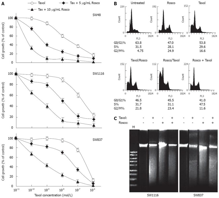
Figure 2 Potentiation of anticancer effect of taxol on human colorectal cancer cell lines by combination with cyclin dependent kinase inhibitor roscovitine.
A: Human colorectal cancer cell lines were treated with taxol (10-11-10-6 mol/L), or the combination of taxol (10-11-10-6 mol/L) and Rosco (5 μg/mL or 10 μg/mL) for 96 h. At the end of treatment, control and drug treated cells were scored for proliferation using the MTT assay; B: Cell cycle phase distribution of human colorectal cancer cells (SW837, 5 × 10⁵ cells/well) treated with taxol (12 × 10-8 mol/L, 72 h); Rosco (15 μg/mL, 72 h); sequential combination: taxol (12 × 10-8 mol/L, 24 h) followed by Rosco (15 μg/mL, 48 h); inverted sequential combination: Rosco (15 μg/mL, 24 h) followed by taxol (12 × 10-8 mol/L, 48 h) and simultaneous combination: taxol plus Rosco (12 × 10-8 mol/L, 15 μg/mL, 72 h) was evaluated by flow cytometry. The percentage of cells in different phases of the cycle was calculated by cell cycle analysis software, Multicycle (Phoenix Flow System, San Diego CA, USA); C: Human colorectal cancer cells, SW1116 and SW837 (5 × 10⁵ cells/well), were treated with taxol (12 × 10-8 mol/L), Rosco (15 μg/mL), and the combination of taxol plus Rosco (12 × 10-8 mol/L + 15 μg/mL) for 72 h. DNA fragments were extracted and analyzed on 1% agrose gel.
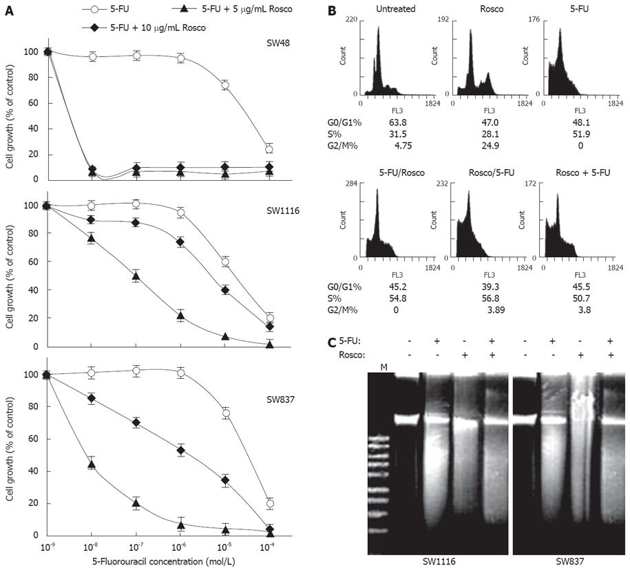
Figure 3 Potentiation of 5-FU anticancer effect on human colorectal cancer cell lines by combination with cyclin dependent kinase inhibitor roscovitine.
A: Human colorectal cancer cell lines were treated with 5-FU (10-9-10-4 mol/L), and the combination of 5-FU (10-9-10-4 mol/L) and Rosco (5 μg/mL or 10 μg/mL) for 96 h. At the end of treatment, control and drug treated cells were scored for proliferation using the MTT assay; B: Cell cycle phase distribution of human colorectal cancer cells (SW837, 5 × 10⁵ cells/well) treated with 5-FU (4.8 × 10⁻⁵ mol/L, 72 h); Rosco (15 μg/mL, 72 h); sequential combination: 5-FU (4.8 × 10⁻⁵ mol/L, 24 h) followed by Rosco (15 μg/mL, 48 h); inverted sequential combination: Rosco (15 μg/mL, 24 h) followed by 5-FU (4.8 × 10⁻⁵ mol/L, 48 h) and simultaneous combination: 5-FU plus Rosco (4.8 × 10⁻⁵ mol/L, 15 μg/mL, 72 h) was evaluated by flow cytometry. The percentage of cells in different phases of the cycle was calculated as described above; C: Human colorectal cancer cells, SW1116 and SW837 (5 × 10⁵ cells/well), were treated with 5-FU (4.8 × 10⁻⁵ mol/L), Rosco (15 μg/L), and the combination of 5-FU plus Rosco (4.8 × 10⁻⁵ mol/L + 15 μg/mL) for 72 h. DNA fragments were extracted and analyzed on 1% agrose gel.
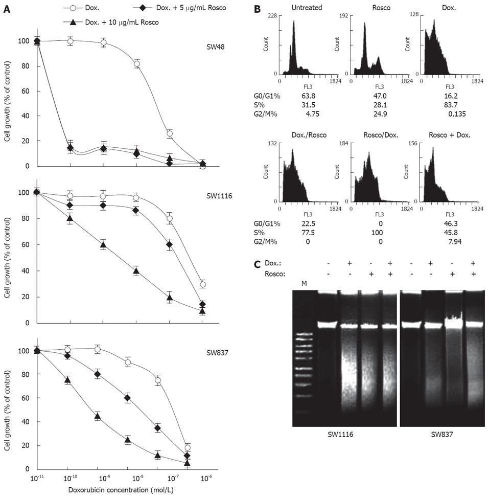
Figure 4 Potentiation of doxorubicin anticancer effect on human colorectal cancer cell lines by combination with cyclin dependent kinase inhibitor roscovitine.
A: Human colorectal cancer cell lines were treated with doxorubicin (10-11-10-6 mol/L), and the combination of doxorubicin (10-11-10-6 mol/L) plus Rosco (5 μg/mL or 10 μg/mL) for 96 h. At the end of treatment, control and drug treated cells were scored for proliferation using an MTT assay; B: Cell cycle phase distribution of human colorectal cancer cells (SW837, 5 × 10⁵ cells/well) treated with doxorubicin (8 × 10⁻⁷ mol/L, 72 h); Rosco (15 μg/mL, 72 h); sequential combination: doxorubicin (8 × 10⁻⁷ mol/L, 24 h) followed by Rosco (15 μg/mL, 48 h); inverted sequential combination: Rosco (15 μg/mL, 24 h) followed by doxorubicin (8 × 10⁻⁷ mol/L, 48 h) and simultaneous combination: doxorubicin plus Rosco (8 × 10⁻⁷ mol/L + 15 μg/mL, 72 h) was evaluated by flow cytometry. The percentage of cells in different phases of the cycle was calculated as previously described; C: Human colorectal cancer cells, SW1116 and SW837, (5 × 10⁵ cells/well) were treated with doxorubicin (8 × 10⁻⁷ mol/L), Rosco (15 μg/mL), and the combination of doxorubicin plus Rosco (8 × 10⁻⁷ mol/L + 15 μg/mL) for 72 h. DNA fragments were extracted and analyzed on 1% agrose gel.
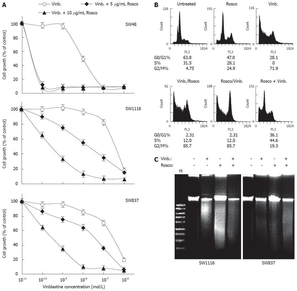
Figure 5 Potentiation of vinblastine anticancer effect on human colorectal cancer cell lines by combination with cyclin dependent kinase inhibitor roscovitine.
A: Human colorectal cancer cell lines were treated with vinblastine (10-12-10-7 mol/L) and the combination of vinblastine (10-12-10-7 mol/L) plus Rosco (5 μg/mL or 10 μg/mL) for 96 h. At the end of treatment, control and drug-treated cells were scored for proliferation using an MTT assay; B: Cell cycle phase distribution of human colorectal cancer cells (SW837, 5 × 10⁵ cells/well) treated with vinblastine (2.6 × 10⁻⁷ mol/L, 72 h); Rosco (15 μg/mL, 72 h); sequential combination: vinblastine (2.6 × 10⁻⁷ mol/L, 24 h) followed by Rosco (15 μg/mL, 48 h); inverted sequential combination: Rosco (15 μg/mL, 24 h) followed by vinblastine (2.6 × 10⁻⁷ mol/L, 48 h) and simultaneous combination: vinblastine plus Rosco (2.6 × 10⁻⁷ mol/L + 15 μg/mL, 72 h) was evaluated by flow cytometry. The percentage of cells in different phases of the cycle was calculated as described above; C: Human colorectal cancer cells, SW1116 and SW837, (5 × 10⁵ cells/well) were treated with vinblastine (2.6 × 10⁻⁷ mol/L), Rosco (15 μg/mL) and the combination of vinblastine plus Rosco (2.6 × 10⁻⁷ mol/L + 15 μg/mL) for 72 h. DNA fragments were extracted and analyzed on 1% agrose gel.
- Citation: Abaza MSI, Bahman AMA, Al-Attiyah RJ. Roscovitine synergizes with conventional chemo-therapeutic drugs to induce efficient apoptosis of human colorectal cancer cells. World J Gastroenterol 2008; 14(33): 5162-5175
- URL: https://www.wjgnet.com/1007-9327/full/v14/i33/5162.htm
- DOI: https://dx.doi.org/10.3748/wjg.14.5162


