Copyright
©2008 The WJG Press and Baishideng.
World J Gastroenterol. Aug 14, 2008; 14(30): 4830-4833
Published online Aug 14, 2008. doi: 10.3748/wjg.14.4830
Published online Aug 14, 2008. doi: 10.3748/wjg.14.4830
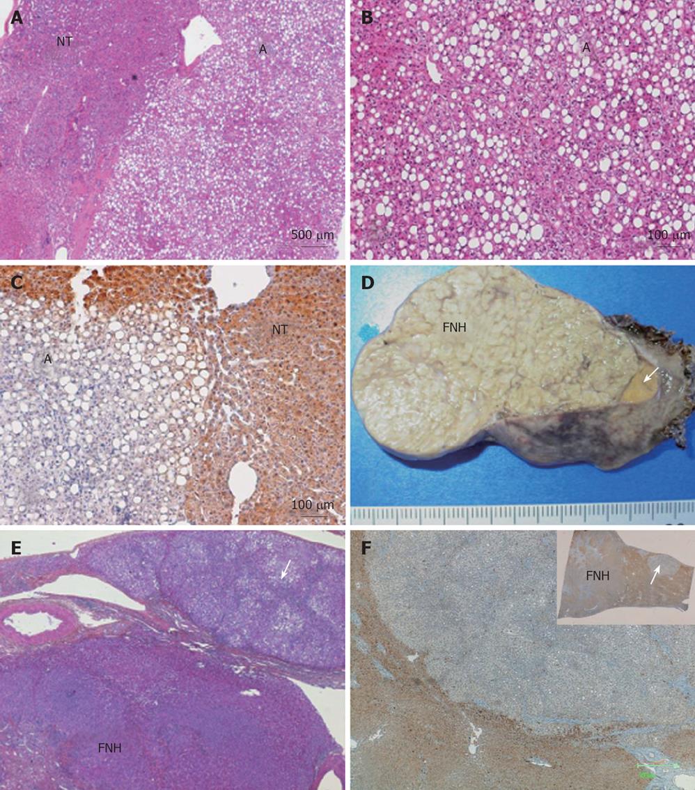
Figure 1 Case 1- HE staining for steatotic adenoma showing non-encapsulated nodule (A, B) and non-tumoral liver (NT) (A), LFABP immunostaining for steatotic or non steatotic tumoral hepatocytes showing no expression of LFABP (C); Case 2- a small yellowish nodule (arrow) adjacent to a typical focal nodular hyperplasia (FNH) (D), HE staining for the small nodule showing a steatotic adenoma adjacent to the FNH (E) and another small steatotic microadenoma (arrows) in the non-tumoral liver (F).
- Citation: Laumonier H, Rullier A, Saric J, Balabaud C, Bioulac-Sage P. Unexpected discovery of 2 cases of hepatocyte nuclear factor 1α-mutated infracentimetric adenomatosis. World J Gastroenterol 2008; 14(30): 4830-4833
- URL: https://www.wjgnet.com/1007-9327/full/v14/i30/4830.htm
- DOI: https://dx.doi.org/10.3748/wjg.14.4830


