Copyright
©2008 The WJG Press and Baishideng.
World J Gastroenterol. Aug 14, 2008; 14(30): 4739-4744
Published online Aug 14, 2008. doi: 10.3748/wjg.14.4739
Published online Aug 14, 2008. doi: 10.3748/wjg.14.4739
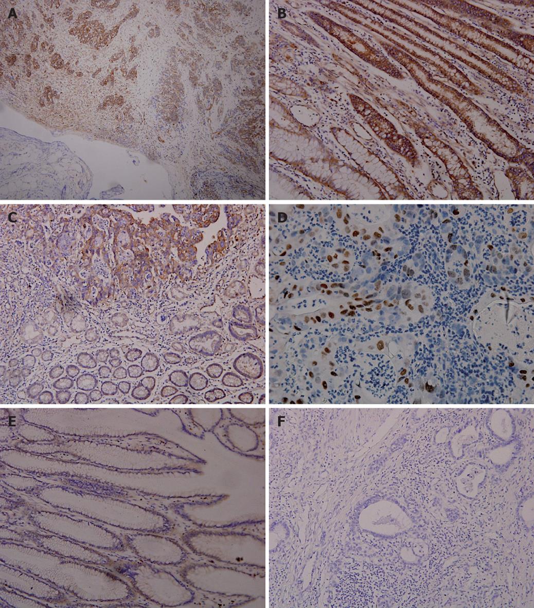
Figure 1 Immunohistochemical staining for NF-κB-p65 showing p65 expression in cancer tissue samples but not in adjacent non-neoplastic tissue samples (SABC, × 100) (A), in gastric adenocarcinoma epithelial tissue samples (SABC, × 200) (B), in intestinal metaplasia samples (SABC, × 200) (C), in nuclei of cancer cells (SABC, × 200) (D), and very weak expression of NF-κB-p65 in gastric mucosa samples (SABC, × 200) (E), and in negative control (SABC, × 200) (F).
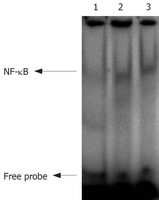
Figure 2 Images of EMSA.
Lanes 1-3: NF-κB activity in normal, moderately- and poorly- differentiated gastric carcinoma tissue samples.
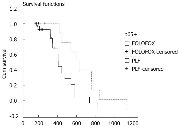
Figure 3 Survival cures for OS time of patients after treatment with PLF and FOLFOX.
- Citation: Ye S, Long YM, Rong J, Xie WR. Nuclear factor kappa B: A marker of chemotherapy for human stage IV gastric carcinoma. World J Gastroenterol 2008; 14(30): 4739-4744
- URL: https://www.wjgnet.com/1007-9327/full/v14/i30/4739.htm
- DOI: https://dx.doi.org/10.3748/wjg.14.4739


