Copyright
©2008 The WJG Press and Baishideng.
World J Gastroenterol. Aug 7, 2008; 14(29): 4690-4693
Published online Aug 7, 2008. doi: 10.3748/wjg.14.4690
Published online Aug 7, 2008. doi: 10.3748/wjg.14.4690
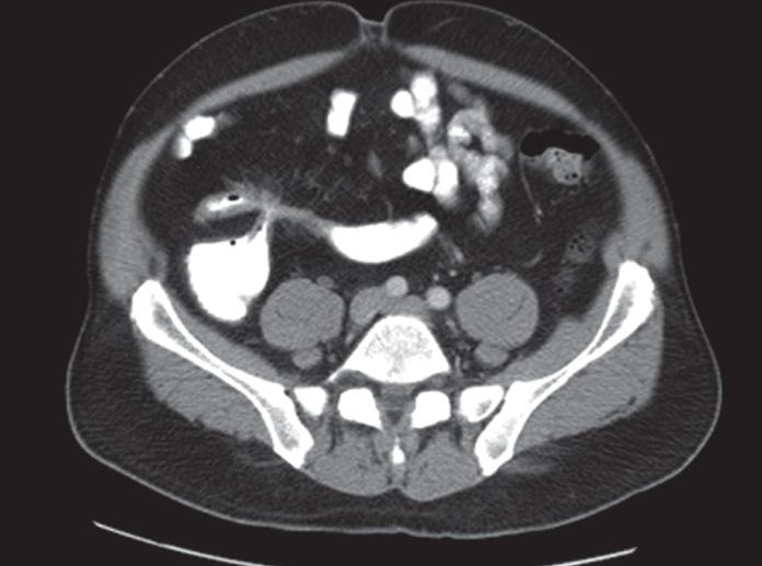
Figure 1 CT scan of the abdomen showing thickened and spiculated ileal wall.
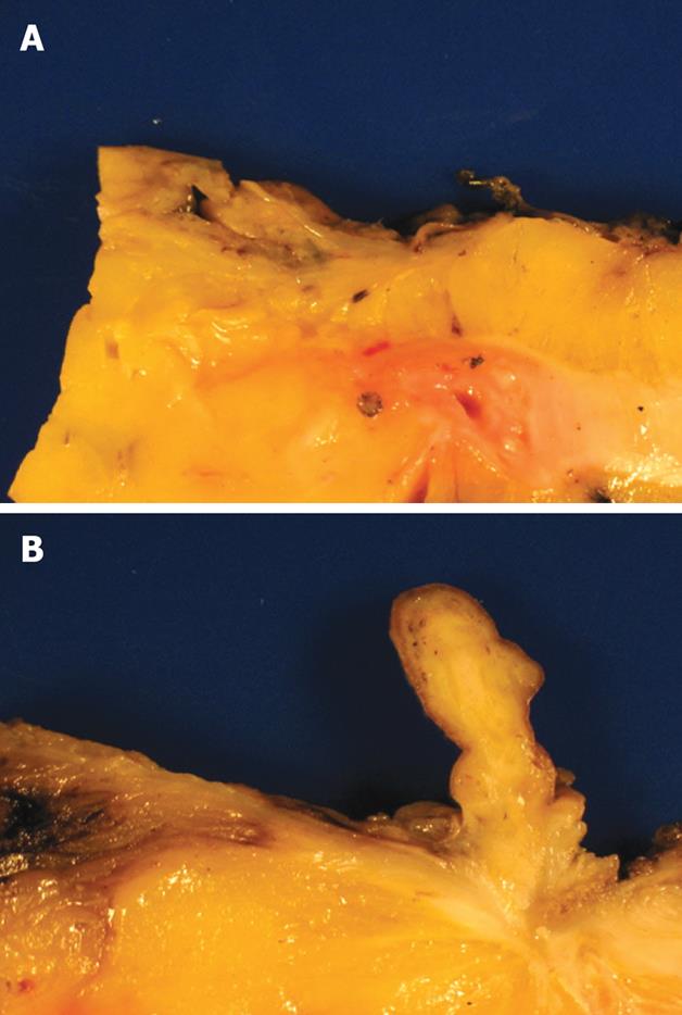
Figure 2 A: Stricture adjacent to the ileocecal valve; B: 3 cm polyp, sections of which revealed a white spiculated area extending into the underlying fat.
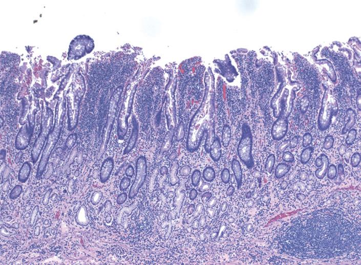
Figure 3 Crohn’s changes in the terminal ileum (HE, × 400).
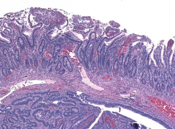
Figure 4 Adenocarcinoma invading the muscularis propria (HE, × 400).
- Citation: Reddy VB, Aslanian H, Suh N, Longo WE. Asymptomatic ileal adenocarcinoma in the setting of undiagnosed Crohn’s disease. World J Gastroenterol 2008; 14(29): 4690-4693
- URL: https://www.wjgnet.com/1007-9327/full/v14/i29/4690.htm
- DOI: https://dx.doi.org/10.3748/wjg.14.4690


